
Membranous Collection
"Exploring the Intricate World Structures: From Mitochondria to Golgi Apparatus" Delving into the Powerhouses


For sale as Licensed Images
Choose your image, Select your licence and Download the media
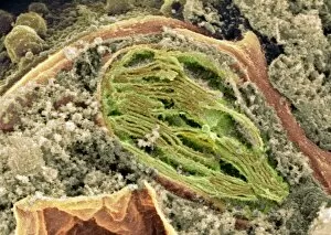
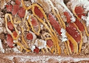
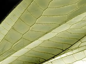
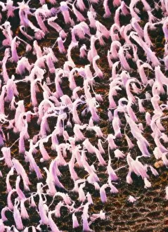
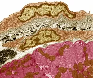
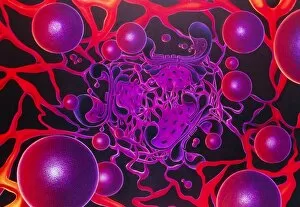
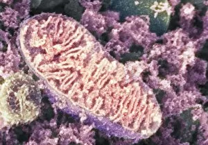
"Exploring the Intricate World Structures: From Mitochondria to Golgi Apparatus" Delving into the Powerhouses: A stunning SEM image reveals the intricate network of mitochondria, highlighting their vital role in cellular energy production. Unveiling Nature's Solar Panels: Witnessing chloroplasts in action through a mesmerizing SEM image, showcasing their ability to convert sunlight into energy during photosynthesis. The Inner Workings Revealed: Another captivating SEM image showcases the detailed structure of mitochondria, providing insights into their crucial functions within cells. Artistic Depiction of Cellular Energy Factories: An exquisite artwork captures the essence and complexity of mitochondrial structures, merging science and art seamlessly. Journeying Through Microscopic Realms: Dive deeper into mitochondrial wonders with artwork C013 / 4993, offering a unique perspective on these essential organelles. Unraveling Mysteries at a Cellular Level: Explore another captivating artwork (C013 / 4991) that beautifully portrays mitochondria's significance in maintaining cellular health and balance. Beauty in Detail: Discover the delicate lacewing wing under an SEM microscope, revealing its intricate membrane structure that aids flight and survival. Peering Into Sound Perception: A false-color SEM unveils macula utriculi within our inner ear - an astonishing sight responsible for detecting motion and gravity changes. Marvels Within Our Hearts' Shield: Observe pericardium tissue at an unprecedented level with TEM imaging, unraveling its protective role around our precious hearts. Captivating Microcosm Revealed Again. Witness yet another breathtaking TEM image showcasing pericardium tissue's remarkable structural features up close. Artistic Glimpse Into Secretive Pathways: An awe-inspiring artwork brings forth the enigmatic beauty of Golgi apparatus – a complex cellular structure involved in protein modification and transport.