
Menisci Collection

Explore the intricate world of knee anatomy with our captivating artwork series

Background imageMenisci Collection: Knee bones and ligaments, artwork C016 / 7012

Knee bones and ligaments, artwork C016 / 7012
Knee bones and ligaments. Artwork of a frontal view of the knee joint, showing bones, ligaments, tendons, and cartilage. The bones are the femur (thigh bone, top), patella (knee-cap, upper centre)

Background imageMenisci Collection: Knee bones and ligaments, artwork C016 / 7011

Knee bones and ligaments, artwork C016 / 7011
Knee bones and ligaments. Artwork of a frontal view of the knee joint, showing bones, ligaments, tendons and cartilage. The bones are the femur (thigh bone, top), patella (knee-cap, upper centre)

Background imageMenisci Collection: Knee anatomy, artwork C016 / 2883

Knee anatomy, artwork C016 / 2883
Knee flexion anatomy. Artwork of a frontal (anterior) view of the bones and some of the cartilage and ligaments of a flexed knee joint

Background imageMenisci Collection: Knee flexion anatomy, artwork C016 / 2880

Knee flexion anatomy, artwork C016 / 2880
Knee flexion anatomy. Artwork of a side (lateral) view of the bones and some of the cartilage and ligaments and tendons of a flexed (left) and extended (right) knee joint

Background imageMenisci Collection: Knee flexion anatomy, artwork C016 / 2881

Knee flexion anatomy, artwork C016 / 2881
Knee flexion anatomy. Artwork of a side (lateral) view of the bones and some of the cartilage and ligaments and tendons of a flexed knee joint

Background imageMenisci Collection: Knee flexion anatomy, artwork C016 / 2879

Knee flexion anatomy, artwork C016 / 2879
Knee flexion anatomy. Artwork of a frontal (anterior) view of the bones and some of the cartilage and ligaments of an extended (left) and flexed (right) knee joint

Background imageMenisci Collection: Knee meniscus tear

Knee meniscus tear
MODEL RELEASED. Knee meniscus tear. Coloured magnetic resonance imaging (MRI) scan of a coronal (front) section through the right knee joint of a 52 year old woman

Background imageMenisci Collection: Rheumatoid arthritis of the knee

Rheumatoid arthritis of the knee joint, artwork. This is a condition in which the bodys immune system attacks the tissues of the joints

Background imageMenisci Collection: Inflamed knee cartilage, computer artwork

Inflamed knee cartilage, computer artwork
Inflamed knee cartilage. Computer artwork of a knee joint, showing the menisci, cartilage discs that lie between the bones providing a smooth surface for movement


For sale as Licensed Images

Choose your image, Select your licence and Download the media

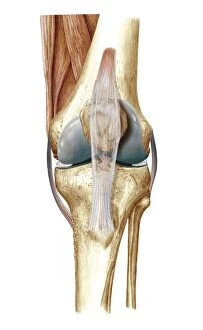
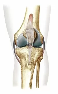
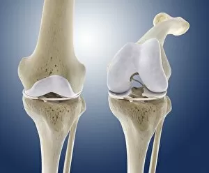
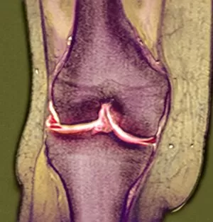
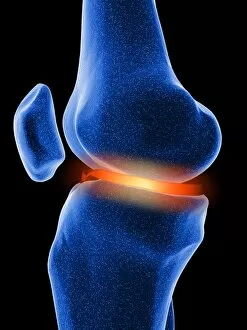
Explore the intricate world of knee anatomy with our captivating artwork series. Witness the crucial roles of menisci, C-shaped cartilages located between the thigh and shin bones (C016 / 7011, C016 / 7012), in knee flexion (C016 / 2880-2881) and stability. However, these vital structures can be susceptible to injuries such as meniscus tears (C016 / 2879, C016 / 2883) or degenerative conditions like rheumatoid arthritis (inflamed knee cartilage, computer artwork). Keep your knees healthy and strong to ensure a pain-free and active lifestyle.