
Metaphase Collection
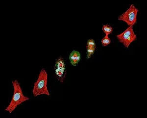
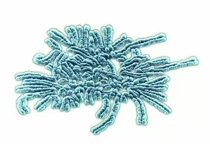
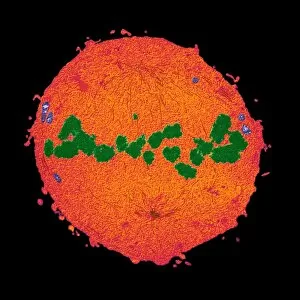
Metaphase, a crucial stage in both mitosis and meiosis, is captured beautifully under the lens of a light micrograph


For sale as Licensed Images
Choose your image, Select your licence and Download the media
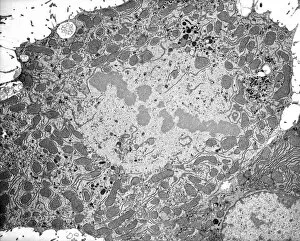
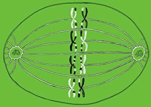
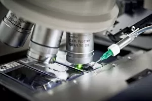
Metaphase, a crucial stage in both mitosis and meiosis, is captured beautifully under the lens of a light micrograph. Dividing cells are seen aligning their chromosomes along the equatorial plate during metaphase, preparing for the subsequent separation. In another stunning image taken with a transmission electron microscope (TEM), we witness the intricate details of a dividing cell during metaphase. The Metaphase II oocyte, as observed through a light micrograph, showcases its readiness for fertilization. Illustration C018 / 0803 depicts the process of meiosis where metaphase plays an essential role in ensuring proper chromosome distribution. Genetic metaphase analysis (C019 / 0289) provides valuable insights into chromosomal abnormalities and genetic disorders by examining dividing cells at this critical phase. Another snapshot from genetic metaphase analysis (C019 / 0288) sheds light on further research conducted to unravel complex genetic patterns. TEM images continue to amaze us with their ability to capture mitosis in action – revealing the intricate machinery that orchestrates cell division during metaphase. Multiple instances of mitotic cells emphasize its significance in various biological processes. Even meiosis gets its moment under scrutiny through scanning electron microscopy (SEM), showcasing how metaphase contributes to gamete formation and genetic diversity.