
Micrograph Collection
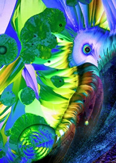
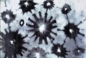
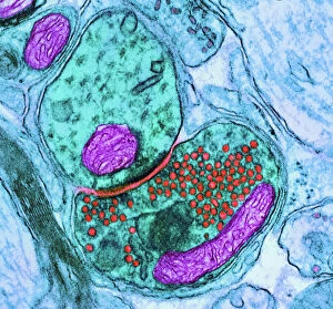
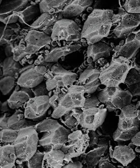
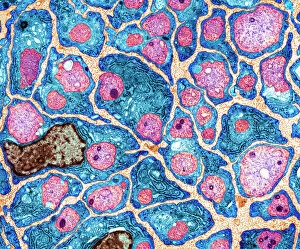
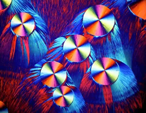
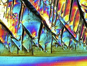
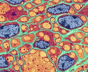
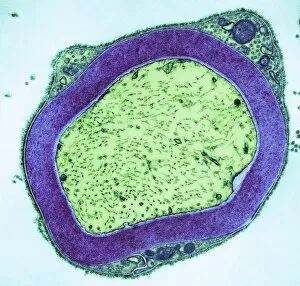
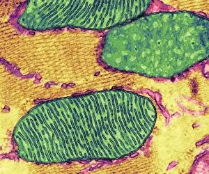
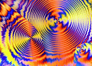
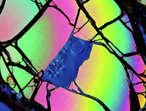
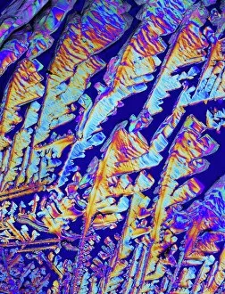
Exploring the intricate world of micrographs: From synapse nerve junctions captured through TEM to the mesmerizing beauty of copper and magnesium sulphate under LM

For sale as Licensed Images
Choose your image, Select your licence and Download the media
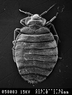
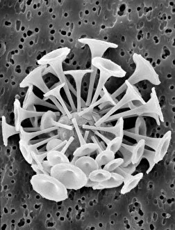
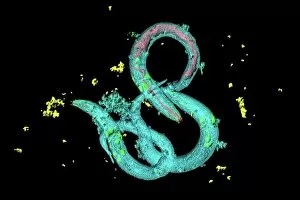
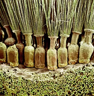
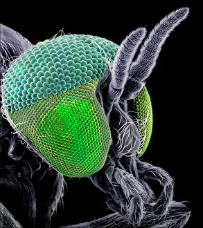
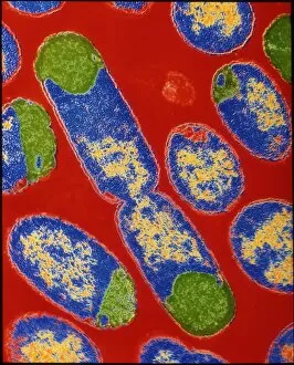
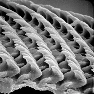
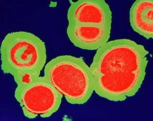
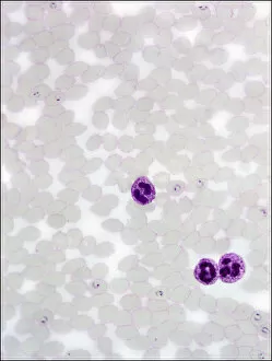
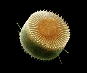
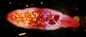
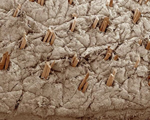
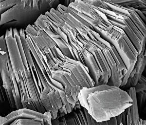
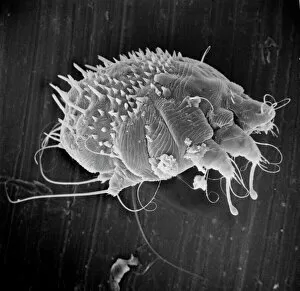
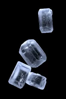
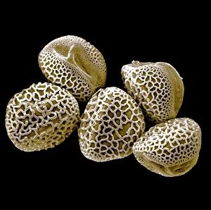
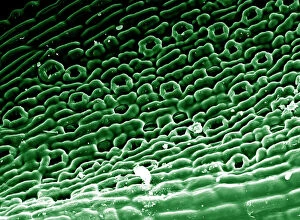
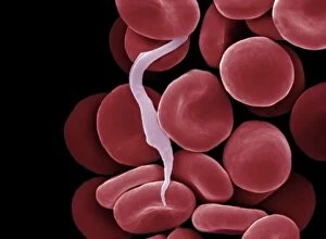
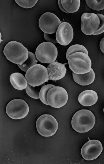
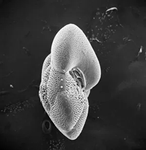
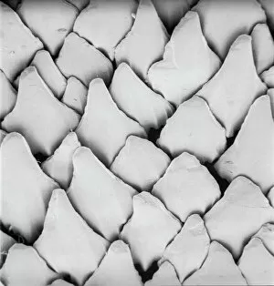
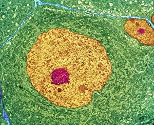
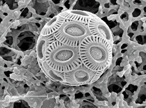
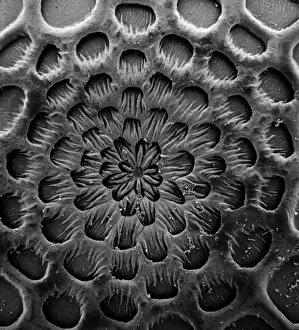
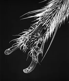
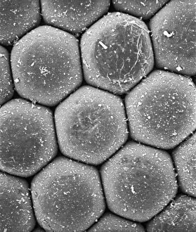
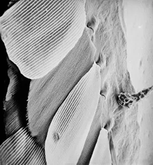
Exploring the intricate world of micrographs: From synapse nerve junctions captured through TEM to the mesmerizing beauty of copper and magnesium sulphate under LM. Witness the delicate structure of Discosphaera tubifera, a coccolithophore, or marvel at the dangerous Crysotile asbestos fibers. Delve into the inner workings of a liver cell or encounter the notorious Cimex lectularius, better known as bed bugs. Admire the ethereal dandelion fruiting head, Taraxacum officinale, or study Simulium damnosum, a Simulian blackfly responsible for river blindness. Uncover E. coli bacteria's microscopic realm or be amazed by snail teeth that defy their size. Behold a stunning silicon crystal in all its glory through light micrography and observe C. elegans worms navigating their environment with grace. These glimpses into unseen worlds remind us of nature's complexity and endless wonders.