
Microorganisms Collection (#6)
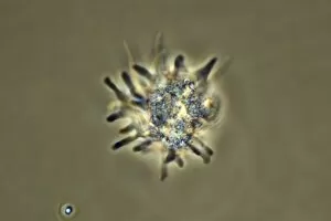
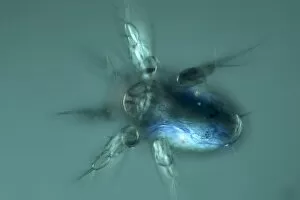
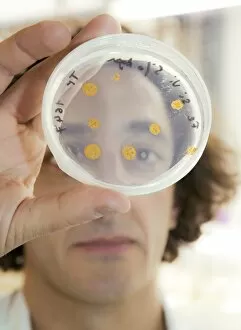
Microorganisms, the tiny wonders of life that exist all around us, are both fascinating and formidable


For sale as Licensed Images
Choose your image, Select your licence and Download the media
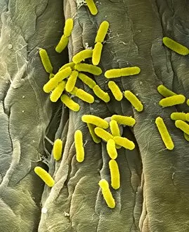
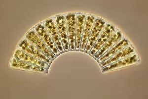
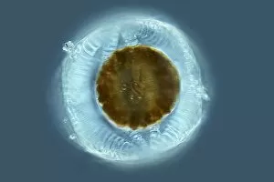
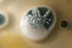
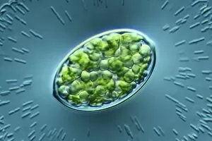
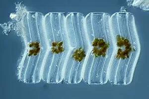
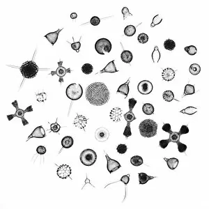
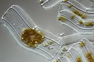
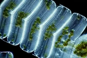
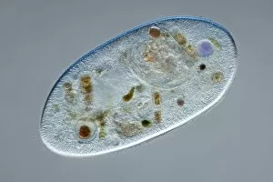
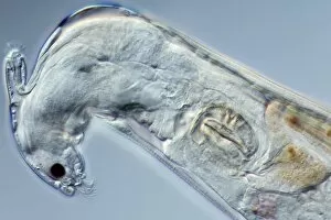
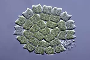
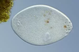
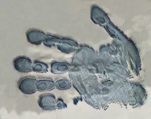
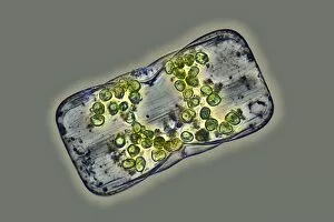
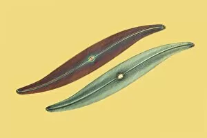
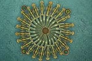
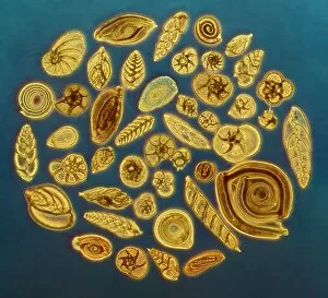
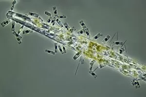
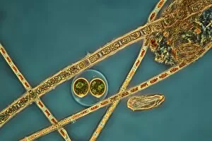
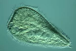
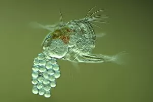
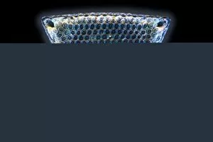
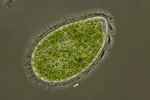
Microorganisms, the tiny wonders of life that exist all around us, are both fascinating and formidable. From the resilient water bear to the intricate structures captured in light micrographs, these microscopic creatures never cease to amaze. Infections spread by sneezing remind us of their potential impact on our health. The artwork depicting this transmission serves as a stark reminder of how easily these unseen organisms can wreak havoc within our bodies. The water bear, observed through scanning electron microscopy, showcases its remarkable resilience and adaptability. Its ability to survive extreme conditions has earned it the title of nature's toughest creature. Looking back at history, lithographs from 1906 reveal colonies of various bacteria that have plagued humanity for centuries. Haemophilus influenzae, Mycobacterium leprae, Micrococcus Gonorrhoea - each with their own unique characteristics and consequences for human health. Streptococcus pneumoniae takes center stage in another lithograph series; its bubble capsule highlighting its role in causing severe respiratory infections. These images serve as a testament to the ongoing battle between humans and disease-causing microorganisms throughout history. Spirochaetes Borrelia Recurrentis is responsible for Lyme disease - an illness that continues to affect countless individuals worldwide. A lithograph showcasing this spirillum highlights the importance of early detection and treatment when dealing with such infectious agents. Vibrio cholerae is yet another notorious bacterium depicted in a lithograph from 1906. This organism causes cholera outbreaks that have claimed countless lives over centuries past and present. Clostridium tetani reveals itself through spores in another captivating lithograph from 1906. This bacterium causes tetanus - a potentially deadly infection often associated with puncture wounds or contaminated objects. Lastly, Mycobacterium tuberculosis reminds us of one of humanity's oldest adversaries: tuberculosis. This ancient pathogen still poses significant challenges today despite medical advancements.