
Microscope Collection (#7)
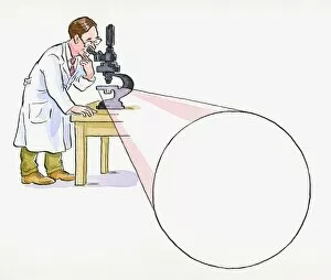
"Unveiling the Unseen: Exploring the Microscopic World" Step into a realm of hidden wonders


For sale as Licensed Images
Choose your image, Select your licence and Download the media
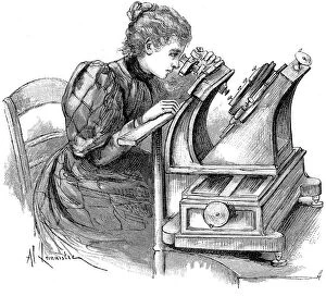
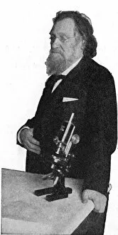
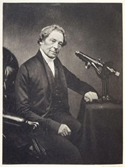
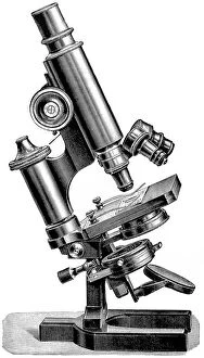
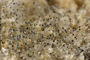
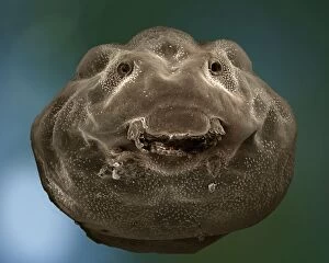
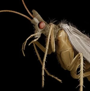
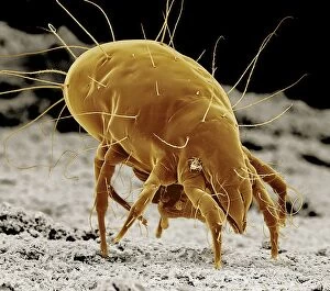
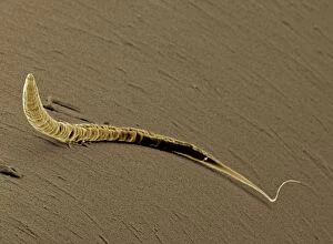
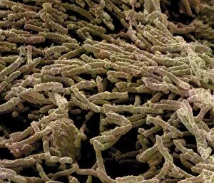
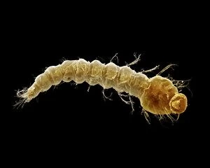
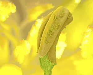
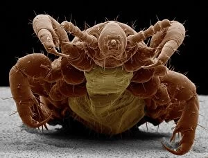
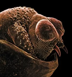
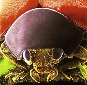
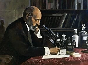
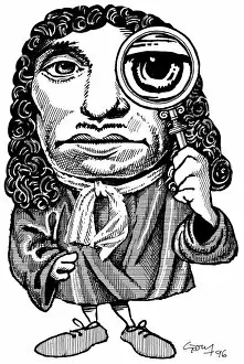
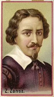
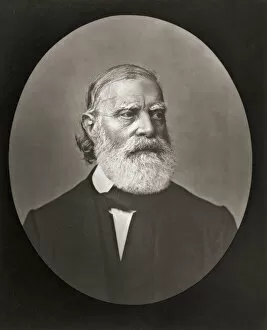
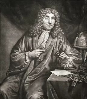
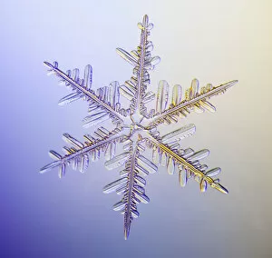
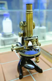
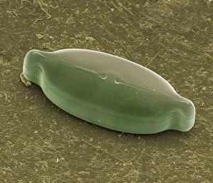
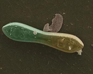
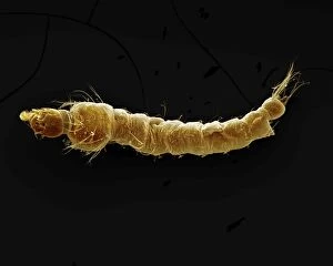
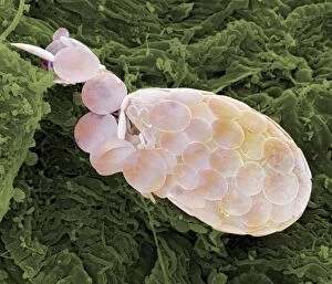
"Unveiling the Unseen: Exploring the Microscopic World" Step into a realm of hidden wonders, where scientists like Rosalind Franklin and Santiago Ramon y Cajal have unraveled the mysteries through their microscopic lens. From Liver Fluke to Norovirus particles, these pioneers have paved the way for groundbreaking discoveries. Witness the intricate beauty of Snail teeth and Fat cells as they come alive under the microscope's gaze. Marvel at Lubbock's Graphic 84, a visual masterpiece capturing the essence of this microcosmic universe. Delve deeper into this microscopic world and encounter Plasmodium sp. , a malarial parasite that has plagued humanity for centuries. Explore E. Coli bacterium in all its glory, revealing both its potential dangers and scientific significance. Through microscope slide preparation techniques, we gain insight into complex organisms like never before. Discover how Fleming used his photographic skills to document crucial moments in microbiology history. The microscope acts as our window into an unseen dimension - it allows us to explore realms beyond our naked eye's reach, and is through this powerful tool that we continue to unravel nature's secrets one magnified image at a time. So join us on this journey "Under the Microscope, " where every slide holds a story waiting to be told – stories that shape our understanding of life itself.