
Microscopes Collection (#2)
Microscopes have revolutionized our understanding of the microscopic world, allowing us to explore and unravel the intricate details of various organisms and structures


For sale as Licensed Images
Choose your image, Select your licence and Download the media
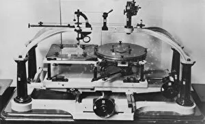
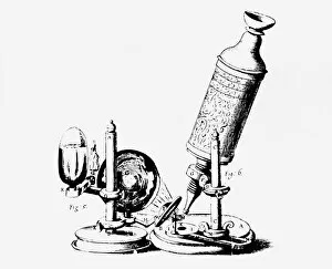
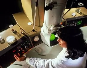
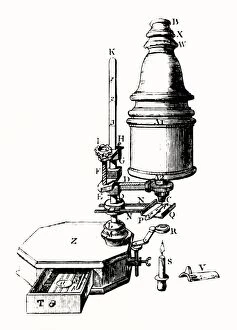
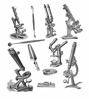
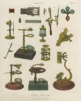
Microscopes have revolutionized our understanding of the microscopic world, allowing us to explore and unravel the intricate details of various organisms and structures. In the realm of parasitology, microscopes play a crucial role in studying organisms like the Liver Fluke. Through meticulous microscope slide preparation techniques, scientists can examine the entire animal under high magnification, revealing its complex anatomy and life cycle. Venturing into nature's hidden treasures, we encounter mesmerizing creatures like the Tardigrade. In a false-coloured SEM image captured amidst moss from Derbyshire's Peak District National Park, this tiny tardigrade appears larger than life. Measuring just 0. 1 millimeters long, it showcases its resilience and beauty through this digital composite. The Thrip's head presents another captivating sight when observed through scanning electron microscopy. The bulging compound eyes on either side reveal an array of sensory hairs sprouting between individual units called ommatidia. This intricate structure highlights nature's remarkable adaptations for visual perception. It also allow us to delve into botanical wonders such as pollen grains found on the style of a Harebell flower at Hoe Grange Nature Reserve in Derbyshire during August. A false-coloured SEM image reveals stunning details that would otherwise remain invisible to our naked eye. Looking back in history, we witness Dr Koch diligently searching for the Rinderpest Microbe at Kimberley while utilizing early optical microscopes in lithographed illustrations from times past. These images remind us of how far microscopy has come since those pioneering days. From advertisements showcasing Stathams combined telescope and microscope to vintage chemistry sets with accompanying microscopes dating back to 1948; these artifacts reflect society's fascination with scientific exploration throughout time. Even artistry finds inspiration in microscopes as seen in beautiful inventions cards featuring chromolitho renderings or trade cards depicting scientific instrument makers like John Smith engraving their legacy onto history.