Microscopic Photos Collection (page 7)

Discovering the hidden wonders of the microscopic world is like entering a realm of infinite fascination

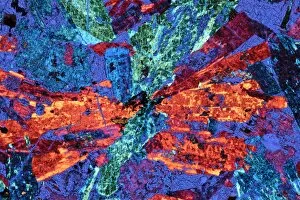
Background imageMicroscopic Photos Collection: Syenite, thin section, polarized LM

Syenite, thin section, polarized LM
Polarised light micrograph of a thin section of syenite, a coarse-grained igneous rock, containing the mineral augite. Augite occurs as a prismatic crystal which in cross-section appears almost

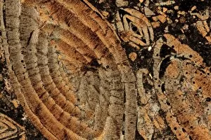
Background imageMicroscopic Photos Collection: Limestone, thin section, polarised LM

Limestone, thin section, polarised LM
Nummulitic limestone. Polarised light micrograph of a thin section of a Nummulitic limestone. The large coloured objects are the remains of large foraminifera, Nummulites sp

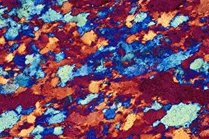
Background imageMicroscopic Photos Collection: Quartzite, thin section, polarized LM

Quartzite, thin section, polarized LM
Polarised light micrograph of a thin section of quartzite, a metamorphic rock. The composition of quartzite varies: quartz is an essential component, with accessories of mica and feldspar

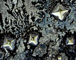
Background imageMicroscopic Photos Collection: Salt crystals, light micrograph

Salt crystals, light micrograph
Salt. Dark field light micrograph of sodium chloride crystals, the common salt. This is a white crystalline compound widespread in nature as a solid (rock salt)

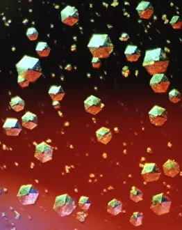
Background imageMicroscopic Photos Collection: Insulin crystals, light micrograph

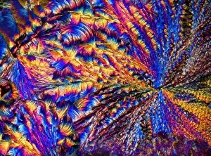
Insulin crystals, light micrograph
Insulin. Polarised light micrograph (PLM) of crystals of the hormone insulin. The crystals appear hexagonal in shape. The insulin molecule is made up of two chains of amino acids (A & B chains)

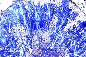
Background imageMicroscopic Photos Collection: Purkinje nerve cells, light micrograph

Purkinje nerve cells, light micrograph
Purkinje nerve cells. Light micrograph of a section through a row of Purkinje nerve cells from the brains cerebellum. Each Purkinje cell is composed of a flask-shaped cell body which branch numerous
Background imageMicroscopic Photos Collection: Silver Nitrate, light micrograph

Silver Nitrate, light micrograph
Composite crystals of Silver Nitrate, polarised light micrograph

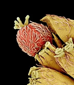
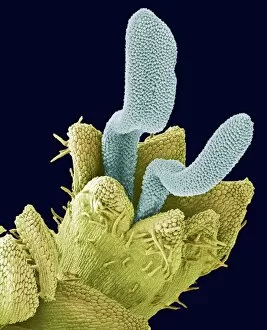
Background imageMicroscopic Photos Collection: Euphorbia flower reproductive parts, SEM

Euphorbia flower reproductive parts, SEM
Euphorbia flower reproductive parts. Coloured scanning electron micrograph (SEM) of the reproductive parts of a Euphorbia supina flower

Background imageMicroscopic Photos Collection: Polarised LM of vitamin D crystals

Polarised LM of vitamin D crystals
Vitamin D. Polarised light micrograph of crystals of vitamin D. Vitamin D is fat-soluble, and is found in foods such as animal fats, milk, butter and eggs

Background imageMicroscopic Photos Collection: Polarised LM of niacinamide crystals

Polarised LM of niacinamide crystals
Nicotinamide. Polarised light micrograph of crystals of nicotinamide, or niacinamide. This is the amide of nicotinic acid. The acid and amide are both B vitamins, and are equally active

Background imageMicroscopic Photos Collection: Treponema pallidum bacteria

Treponema pallidum bacteria

Background imageMicroscopic Photos Collection: Gas gangrene bacteria, SEM

Gas gangrene bacteria, SEM
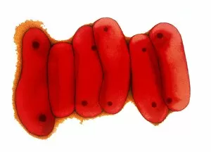
Gas gangrene bacteria (Clostridium perfringens), coloured scanning electron micrograph (SEM). These rod-shaped bacteria are pathogenic

Background imageMicroscopic Photos Collection: Tuberculosis bacteria, TEM

Tuberculosis bacteria, TEM
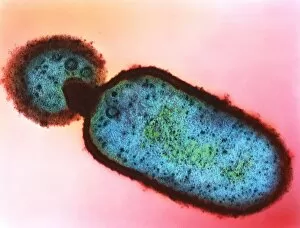
Tuberculosis bacteria. Coloured transmission electron micrograph (TEM) of a section through Mycobacterium tuberculosis bacteria

Background imageMicroscopic Photos Collection: Moraxella catarrhalis bacteria

Moraxella catarrhalis bacteria

Background imageMicroscopic Photos Collection: Rhodopseudomonas sp bacteria

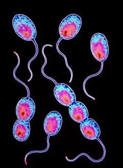
Rhodopseudomonas sp bacteria

Background imageMicroscopic Photos Collection: SEM of soil bacteria

SEM of soil bacteria

Background imageMicroscopic Photos Collection: Penicillin fungus growing on cheddar cheese

Penicillin fungus growing on cheddar cheese
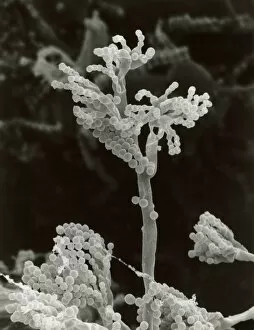
Scanning electron micrograph (SEM) the fruiting bodies or conidia of a penicillin fungus growing on a piece of English cheddar cheese which has gone mouldy in the refrigerator

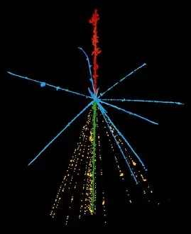
Background imageMicroscopic Photos Collection: Emulsion photo of cosmic ray event

Emulsion photo of cosmic ray event
The power of a cosmic ray is shown in this false- colour emulsion photo of a cosmic ray sulphur nucleus (red) colliding with a nucleus in the emulsion

Background imageMicroscopic Photos Collection: Particle tracks on a star chart

Particle tracks on a star chart

Background imageMicroscopic Photos Collection: Disc floret, SEM

Disc floret, SEM
Disc floret. Coloured scanning electron micrograph (SEM) of a disc floret of a composite flower. Daisies (family Asteraceae, formerly Compositae) have inflorescences composed of two types of flowers

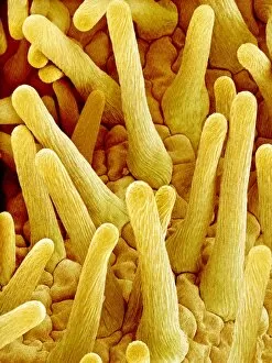
Background imageMicroscopic Photos Collection: Comfrey flower petal, SEM

Comfrey flower petal, SEM
Comfrey flower petal. Coloured scanning electron micrograph (SEM) showing papillae on a petal from a comfrey (Symphytum officinale) flower. Papillae are projections from epidermal cells

Background imageMicroscopic Photos Collection: Open stoma, SEM

Open stoma, SEM
Open stoma. Coloured scanning electron micrograph (SEM) of an open stoma on the surface of a tobacco leaf (Nicotiana tabacum)

Background imageMicroscopic Photos Collection: Light micrograph of vitamin B6 crystals

Light micrograph of vitamin B6 crystals
Polarised light micrograph of crystals of vitamin B6, known as Pyridoxine. Magnification: x63 at 35mm size

Background imageMicroscopic Photos Collection: Clostridium tetani bacterium

Clostridium tetani bacterium
False-colour transmission electron micrograph (TEM) of the bacterium Clostridium tetani, a Gram- positive, spore-forming, anaerobic bacillus (rod-shaped bacterium) that causes tetanus

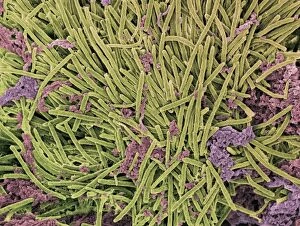
Background imageMicroscopic Photos Collection: Tongue bacteria, SEM

Tongue bacteria, SEM
Tongue bacteria. Coloured scanning electron micrograph (SEM) of bacteria on the surface of a human tongue. Large numbers of bacteria can form a visible layer on the surface of the tongue

Background imageMicroscopic Photos Collection: Illustration of structures of a typical bacterium

Illustration of structures of a typical bacterium
Cutaway illustration of the internal structure of a typical bacterial cell. This example is of a capsulated bacterium; the capsule (light green) forms the outer of 3 layers

Background imageMicroscopic Photos Collection: Tobacco plant protoplast, SEM

Tobacco plant protoplast, SEM
Protoplast. Coloured scanning electron micrograph (SEM) of a tobacco plant (Nicotiana tabacum) protoplast. A protoplast is a plant cell that has had its tough outer cell wall removed by chemical

Background imageMicroscopic Photos Collection: Quantum tunneling

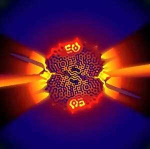
Quantum tunneling. Computer model of a quantum wavefunction trapped in a deep well (centre). In classical physics, the particle described by this wavefunction doesn t have enough energy to emerge

Background imageMicroscopic Photos Collection: Female sex hormone

Female sex hormone. Polarised light micrograph of crystals of Oestradiol, also known as beta- Estradiol. This is the most potent of the six naturally-occurring oestrogens

Background imageMicroscopic Photos Collection: MRSA bacteria, TEM

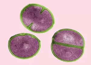
MRSA bacteria, TEM
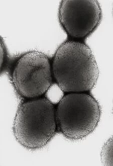
MRSA bacteria. Coloured transmission electron micrograph (TEM) of methicillin-resistant Staphylococcus aureus (MRSA), a gram-positive, round (coccus) bacterium

Background imageMicroscopic Photos Collection: Silver nitrate crystals, LM

Silver nitrate crystals, LM
Silver nitrate crystals. Polarised light micrograph (LM) of silver nitrate (AgNO3) crystals. Silver nitrate is used in photography and as a test for chloride, bromide and iodide ions

Background imageMicroscopic Photos Collection: Magnesium chloride crystals, LM

Magnesium chloride crystals, LM
Magnesium chloride. Polarised light micrograph (LM) of crystals of magnesium chloride (MgCl2). Typically, magnesium chloride forms complexes with other molecules; the hygroscopic hexahydrate

Background imageMicroscopic Photos Collection: Magnesium sulphate crystals, LM

Magnesium sulphate crystals, LM
Magnesium sulphate. Polarised light micrograph of crystals of magnesium sulphate (MgSO4), a chemical used medicinally and in the textile industry

Background imageMicroscopic Photos Collection: Sperm on egg during fertilisation

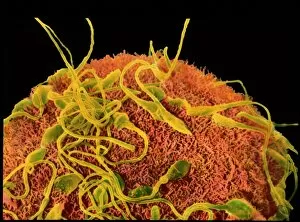
Sperm on egg during fertilisation
Fertilisation. Coloured scanning electron micro- graph (SEM) of sperm on the surface of a human egg (ovum) during fertilisation. The egg has a furry surface (brown)

Background imageMicroscopic Photos Collection: Staphylococcus aureus dividing, TEM

Staphylococcus aureus dividing, TEM
Staphylococcus aureus bacteria dividing, coloured transmission electron micrograph. Atlower left is a single bacterium, the bacterium atupper centre is starting to divide

Background imageMicroscopic Photos Collection: Oral bacteria, TEM

Oral bacteria, TEM
Oral bacteria. Computer-enhanced colouredtransmission electron micrograph (TEM) of Porphyromonas gingivalis bacteria (formerlynamed Bacteroides gingivalis )

Background imageMicroscopic Photos Collection: Bacterial cell

Bacterial cell, computer artwork. This is ageneralised bacterium, illustrating features ofdifferent bacterial types that would not becombined in a single cell

Background imageMicroscopic Photos Collection: Helicobacter pylori bacteria in stomach

Helicobacter pylori bacteria in stomach
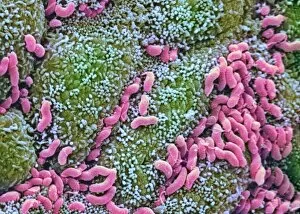
Helicobacter pylori bacteria. Colouredscanning electron micrograph (SEM) of Helicobacter pylori bacteria (pink), a causeof gastritis (stomach lining inflammation)

Background imageMicroscopic Photos Collection: Listeria monocytogenes bacteria, TEM

Listeria monocytogenes bacteria, TEM
Listeria bacteria. Coloured transmissionelectron micrograph (TEM) of a Listeria monocytogenes bacterium (centre to bottomright). It is dividing by a process of unequalcell growth known as budding

Background imageMicroscopic Photos Collection: Unidentified rod-shaped bacteria

Unidentified rod-shaped bacteria
Bacteria. Coloured scanning electronmicrograph (SEM) of unidentified bacilli(rod-shaped bacteria). Magnification unknown


For sale as Licensed Images

Choose your image, Select your licence and Download the media

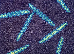
Discovering the hidden wonders of the microscopic world is like entering a realm of infinite fascination. From particle tracks leaving trails of mystery to the mesmerizing bubble chamber photo capturing the decay of a sigma particle, these microscopic photos unveil secrets that lie beyond our naked eye's reach. One remarkable image showcases the first observation of an omega-minus particle, a groundbreaking moment in scientific history. Meanwhile, another snapshot reveals the delicate beauty of a human blastocyst, reminding us of life's miraculous beginnings. Immunofluorescent LM offers us glimpses into intricate networks within our own bodies - neurons and astrocytes intricately woven together like cosmic constellations. And then there are oxytocin hormone crystals, their vibrant colors shining through as if encapsulating love itself. The iris of an eye captured by SEM reminds us that even something as ordinary as our eyes holds extraordinary complexity when seen up close. Similarly, osteoporotic bone under SEM unveils its fragile structure with astonishing detail. Light micrographs bring forth stunning images such as oxytocin crystals glistening like precious gems or spiral spore chains formed by Streptomyces bacteria resembling ethereal works of art. And let us not forget about our retina - a gateway to vision and perception - or nerve cells firing messages at lightning speed throughout our bodies, orchestrating every movement and thought we have. These microscopic photos offer more than just visual delight; they invite us to ponder upon the vastness contained within each minuscule frame. They remind us that sometimes it is in exploring the smallest details that we gain profound insights into ourselves and the world around us.