
Microscopic Collection (#11)
"Exploring the Microscopic World: A Glimpse into Nature's Hidden Wonders" Step into a realm unseen by the naked eye


For sale as Licensed Images
Choose your image, Select your licence and Download the media
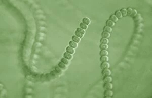
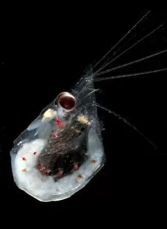
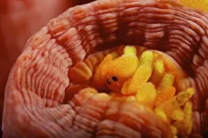
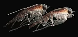
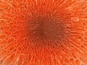
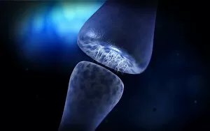
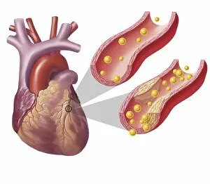
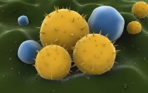
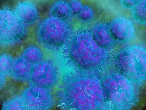
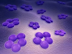
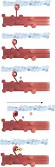
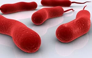
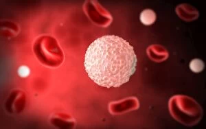
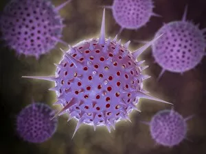
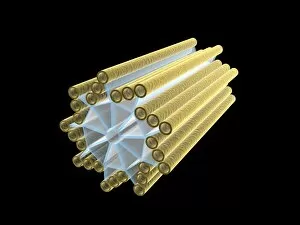
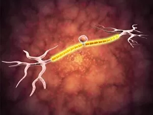
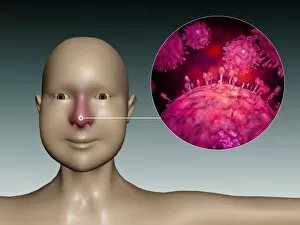
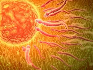
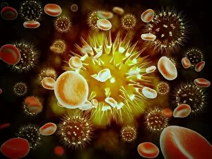
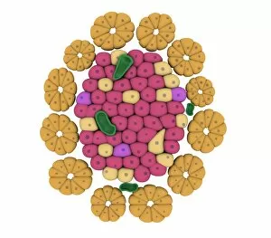
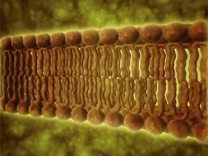
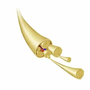
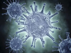
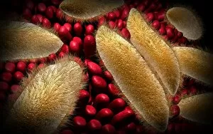
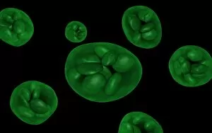
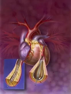
"Exploring the Microscopic World: A Glimpse into Nature's Hidden Wonders" Step into a realm unseen by the naked eye, where tiny creatures and intricate structures come to life. In Picture No. 10851646, we are captivated by the Scanning Electron Micrograph (SEM) of a Praying Mantis, magnified x30. Its delicate features and mesmerizing details reveal a world within our reach. Moving on to another fascinating creature, the Tardigrade or 'Water Bear', magnified x1250 in an SEM image. This resilient micro-animal showcases its remarkable adaptability under extreme conditions. It reminds us that even in the tiniest forms of life, strength can be found. The Fruit Fly takes center stage next with an SEM image at x300 magnification. Despite its minuscule size, this common insect holds secrets waiting to be discovered under closer inspection. Its intricate body structure hints at its role in nature's grand tapestry. Diatoms from marine plankton samples grace our vision next - their elegant shapes and patterns resembling miniature works of art created by nature itself. These microscopic organisms play a vital role in Earth's ecosystems as primary producers. An illustration of a Flea C017/3435 introduces us to yet another hidden world beneath our feet - one filled with parasites and symbiotic relationships that shape entire ecosystems on both macro and micro scales. Diving deeper into marine diatoms at x25 magnification reveals their stunning beauty – intricate designs etched onto tiny shells floating through vast oceans, silently contributing to Earth's biodiversity. Anton van Leeuwenhoek's observations of Animalcules circa 1795 remind us of humanity's earliest encounters with microscopic lifeforms – pioneering discoveries that forever changed our understanding of biology and paved the way for modern scientific advancements. A Macro Photograph unveils Paenibacillus bacteria colonies thriving amidst laboratory-imposed stresses, showcasing their ability to adapt and survive in challenging environments.