Molecules Collection (#34)

"Molecules

Background imageMolecules Collection: Human rhinovirus capsid proteins

Human rhinovirus capsid proteins, molecular model. These are proteins from the capsid (outer protein coat) of rhinovirus 14. Rhinoviruses are responsible for causing about 50% of common colds

Background imageMolecules Collection: Haemagglutinin from bird flu virus

Haemagglutinin from bird flu virus, molecular model. This protein, H5, is found on the surface of the bird flu virus H5N1

Background imageMolecules Collection: Cytosine molecule

Cytosine molecule
Cytosine. Molecular model of the nucleobase cytosine (2-oxy-4-aminopyrimidine). This is a pyrimidine-derived nucleobase found in the genetic molecules DNA (deoxyribonucleic acid)

Background imageMolecules Collection: Alpha-endorphin molecule

Alpha-endorphin molecule. Molecular model of the analgesic (painkilling) peptide alpha-endorphin. This molecule is released by the pituitary gland at times of stress or great pain

Background imageMolecules Collection: Valine, molecular model

Valine, molecular model
Valine. Molecular model of the amino acid valine. Its chemical formula is C5.H11.N.O2. Atoms are represented as balls and are colour-coded: carbon (blue), hydrogen (gold)

Background imageMolecules Collection: Tyrosine, molecular model

Tyrosine, molecular model
Tyrosine. Molecular model of the amino acid tyrosine. Its chemical formula is C9.H11.N.O3. Atoms are represented as rods and are colour- coded: carbon (blue), hydrogen (gold)

Background imageMolecules Collection: Tryptophan, molecular model

Tryptophan, molecular model
Tryptophan. Molecular model of the amino acid tryptophan. Its chemical formula is C11.H12.N2.O2. Atoms are represented as spheres and are colour- coded: carbon (blue), hydrogen (gold)

Background imageMolecules Collection: Threonine, molecular model

Threonine, molecular model
Threonine. Molecular model of the amino acid threonine. Its chemical formula is C4.H9.N.O3. Atoms are represented as spheres and are colour- coded: carbon (blue), hydrogen (gold)

Background imageMolecules Collection: Serine, molecular model

Serine, molecular model
Serine. Molecular model of the amino acid serine. Its chemical formula is C3.H7.N.O3. Atoms are represented as spheres and are colour-coded: carbon (blue), hydrogen (gold)

Background imageMolecules Collection: Pyrrolysine, molecular model

Pyrrolysine, molecular model
Pyrrolysine. Molecular model of the amino acid pyrrolysine. Its chemical formula is C12.H21.N3.O3. Atoms are represented as rods and are colour- coded: carbon (blue), hydrogen (gold)

Background imageMolecules Collection: Proline, molecular model

Proline, molecular model
Proline. Molecular model of the amino acid proline. Its chemical formula is C5.H9.N.O2. Atoms are represented as spheres and are colour- coded: carbon (blue), hydrogen (gold)

Background imageMolecules Collection: Phenylalanine, molecular model

Phenylalanine, molecular model
Phenylalanine. Molecular model of the amino acid phenylalanine. Its chemical formula is C9.H11.N.O2. Atoms are represented as spheres and are colour-coded: carbon (blue), hydrogen (gold)

Background imageMolecules Collection: Methionine, molecular model

Methionine, molecular model
Methionine. Molecular model of the amino acid methionine. Its chemical formula is C5.H11.N.O2.S. Atoms are represented as rods and are colour- coded: carbon (blue), hydrogen (gold)

Background imageMolecules Collection: Leucine, molecular model

Leucine, molecular model
Leucine. Molecular model of the amino acid leucine. Its chemical formula is C6.H13.N.O2. Atoms are represented as spheres and are colour- coded: carbon (blue), hydrogen (gold)

Background imageMolecules Collection: Lysine, molecular model

Lysine, molecular model
Lysine. Molecular model of the amino acid lysine. Its chemical formula is C6.H14.N2.O2. Atoms are represented as spheres and are colour- coded: carbon (blue), hydrogen (gold)

Background imageMolecules Collection: FK506-binding protein molecule

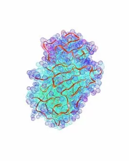
FK506-binding protein molecule. Computer model showing the primary (rods) and secondary (alpha- helices, blue, and beta-sheets)

Background imageMolecules Collection: Lipase molecule

Lipase molecule. Computer model showing the secondary structure of lipase. Alpha-helices are blue and beta-sheets are purple

Background imageMolecules Collection: DNA polymerase Klenow fragment

DNA polymerase Klenow fragment
Klenow fragment of DNA polymerase I. Computer model showing the secondary (alpha-helices and beta-sheets) and primary (ball-and-stick) structures of the Klenow, or large

Background imageMolecules Collection: Protein tyrosine phosphatase molecule

Protein tyrosine phosphatase molecule. Computer model of the secondary structure of an intermediate form of protein tyrosine phosphatase. Beta-sheets are purple and alpha-helices are blue

Background imageMolecules Collection: Cyclin-dependent kinase 2 enzyme

Cyclin-dependent kinase 2 enzyme, molecular model. This enzyme is found in cells, where it is involved in regulating the cell cycle, the cycle of cell division and cell growth

Background imageMolecules Collection: ATPase muscle enzyme

ATPase muscle enzyme
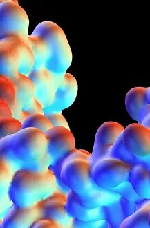
Calcium pumping ATPase enzyme. Computer model of an electrostatic potential surface map of part of the ATPase enzyme that pumps calcium in and out of muscle cells and controls muscle contractions

Background imageMolecules Collection: Pepsin molecule

Pepsin molecule
Pepsin enzyme. Computer graphic of the protein- digesting enzyme pepsin. It is a protease enzyme that is secreted as part of gastric juice into the stomach in an inactive form known as pepsinogen

Background imageMolecules Collection: Fibroblast growth factor receptor 2

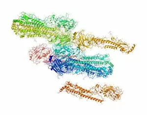
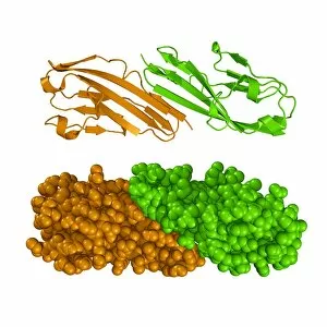
Fibroblast growth factor receptor 2 (FGFR2). Molecular models of the secondary structure (top) and the tertiary structure (bottom) of FGFR2

Background imageMolecules Collection: Enzyme catalysing DNA recombination

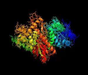
Enzyme catalysing DNA recombination. Computer model of the enzyme flippase recombinase (FLP recombinase, atoms represented as tubes)

Background imageMolecules Collection: Cholesterol, molecular model

Cholesterol, molecular model
Cholesterol. Molecular model of the fatty and waxy alcohol cholesterol. Atoms are represented by spheres and are colour-coded: carbon (black), hydrogen (grey), oxygen (red)


For sale as Licensed Images

Choose your image, Select your licence and Download the media

"Molecules: The Building Blocks of Life and Beyond" From the intricate workings of an anaesthetic inhibiting an ion channel C015/6718 to the genius mind of James Clerk Maxwell, they have captivated scientists and artists alike. With their diverse structures and functions, they hold the key to understanding life at its core. Delving into the world of proteins, we witness their secondary structure through mesmerizing artwork that unveils their complexity. Meanwhile, the caffeine drug molecule keeps us awake while bacterial ribosomes tirelessly synthesize proteins within our cells. Vitamin B12's molecular model reminds us of nature's intricate design as zinc fingers elegantly bind to a DNA strand, orchestrating genetic processes. And who can forget capsaicin - the fiery molecule responsible for giving chili peppers their spicy kick? But molecules aren't limited to just earthly matters; they extend beyond our planet's boundaries. Oxytocin neurotransmitter molecules remind us of love's chemical connection while praziquantel parasite drugs combat infections in distant lands. Interferon molecules stand tall as defenders against viral invasions, showcasing our body's remarkable defense mechanisms. And amidst all this scientific wonder lies a breathtaking sight - Aurora Borealis dancing over a snow-covered coniferous forest in Northern Finland. Intricate and awe-inspiring, these glimpses into the molecular world remind us that there is so much more than meets the eye. From unlocking medical breakthroughs to unraveling nature's mysteries or simply marveling at captivating artistry – they can truly extraordinary entities shaping our understanding of life itself.