Mucosal Collection (#2)

"Mucosal: Unveiling the Intricate World of Our Body's Protective Lining" Our body is a complex system, and within it lies an intricate network known as mucosal lining

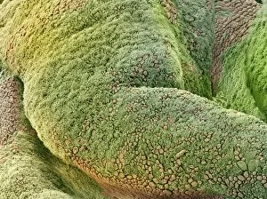
Background imageMucosal Collection: Fallopian tube lining, SEM

Fallopian tube lining, SEM
Fallopian tube lining. Coloured scanning electron micrograph (SEM) of the folded internal mucosal lining of a fallopian tube (oviduct)

Background imageMucosal Collection: Inflamed gall bladder, light micrograph

Inflamed gall bladder, light micrograph
Inflamed gall bladder. Light micrograph of a section through a gall bladder with cholecystitis (inflammation). The mucosa lining (purple) is thickened

Background imageMucosal Collection: Cryptosporidiosis, TEM

Cryptosporidiosis, TEM

Background imageMucosal Collection: Trachea mucous membrane, SEM

Trachea mucous membrane, SEM
Trachea mucous membrane. Coloured scanning electron micrograph (SEM) of a fractured mucous membrane of the trachea (wind pipe), showing the epithelium and underlying connective tissue

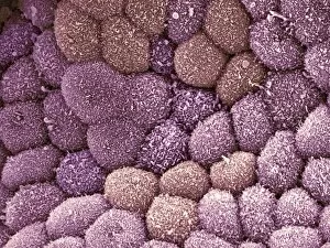
Background imageMucosal Collection: Gall bladder surface, SEM

Gall bladder surface, SEM
Internal lining of the gall bladder, coloured scanning electron micrograph (SEM). These columnar epithelial cells are covered in microvilli. Magnification: x2100 when printed at 10 centimetres wide

Background imageMucosal Collection: Sperm cell, SEM

Sperm cell, SEM
Sperm cell. Coloured scanning electron micrograph (SEM) of a sperm cell in a fallopian tube. Magnification: x400 when printed at 10 centimetres wide

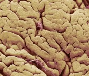
Background imageMucosal Collection: Snake intestinal villi, SEM

Snake intestinal villi, SEM
Snake intestine. Coloured scanning electron micrograph (SEM) of villi in the intestine of a snake. The villi (folds) in the surface of the intestine are short and covered in cuboidal mucosal cells

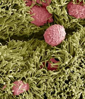
Background imageMucosal Collection: Snake ciliated lung cells and mucus, SEM

Snake ciliated lung cells and mucus, SEM
Snake ciliated lung cells and mucus. Coloured scanning electron micrograph (SEM) of ciliated cells (green) and mucus (pink) in the lung of a snake

Background imageMucosal Collection: Oviduct mucosal folds, light micrograph

Oviduct mucosal folds, light micrograph. This section through the folds shows the ciliated columnar epithelium (red) and the connective tissue core (green)


For sale as Licensed Images

Choose your image, Select your licence and Download the media

"Mucosal: Unveiling the Intricate World of Our Body's Protective Lining" Our body is a complex system, and within it lies an intricate network known as mucosal lining. This remarkable barrier plays a vital role in protecting various organs and tissues from harm. Let's delve into some fascinating glimpses of different mucosal regions: Gastric Antral Vascular Ectasia (GAVE) C016 / 8328: Explore the mesmerizing patterns formed by blood vessels in the stomach's antrum region, highlighting GAVE. Trachea Lining, SEM C013 / 7126: Witness the microscopic beauty of tracheal lining through scanning electron microscopy, revealing its delicate structure. SEM C013 / 7122: Another captivating view showcasing the intricacies of our respiratory system's protective layer - the tracheal lining. Ulcerative Pancolitis C018 / 0928: Observe how inflammation affects the entire colon with this image depicting ulcerative pancolitis, emphasizing mucosal damage. 5-7. Gastritis Light Micrograph (x3): Journey into gastric troubles with these light micrographs capturing gastritis - inflammation of stomach lining caused by various factors. 8-9. Colitis Light Micrograph (x2): Delve deeper into colonic health as these images portray colitis - inflammation affecting large intestine mucosa leading to discomfort. Ischaemic Bowel Light Micrograph: Witness how insufficient blood supply can impact bowel health through this enlightening light micrograph showcasing ischaemic bowel condition. 11-12. Vaginal Mucosa Light Micrograph (x2): Discover the importance of vaginal mucosa in maintaining reproductive health through these vivid light micrographs that unveil its unique characteristics.