
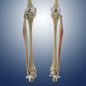
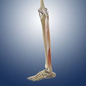
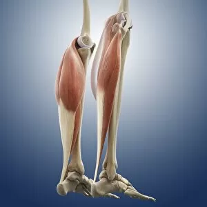
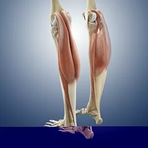
Muscular System Collection (#5)

"The Marvels of the Muscular System

Background imageMuscular System Collection: Human muscle fibres, diagram

Human muscle fibres, diagram
Human muscle fibres. Diagram showing the structure of human muscles from the cellular to the macroscopic level. At lower right, muscle cells contain many myofibrils

Background imageMuscular System Collection: Neck muscle, artwork C014 / 5019

Neck muscle, artwork C014 / 5019
Neck muscle. Computer artwork showing the splenius capitis muscle of the neck. This muscle is attached to the upper thoracic (chest) and lower cervical (neck) vertebre and the base of the skull

Background imageMuscular System Collection: Neck muscle, artwork C014 / 5022

Neck muscle, artwork C014 / 5022
Neck muscle. Computer artwork showing the splenius capitis muscle of the neck. This muscle is attached to the upper thoracic (chest) and lower cervical (neck) vertebre and the base of the skull

Background imageMuscular System Collection: Deltoid muscle, artwork C014 / 5010

Deltoid muscle, artwork C014 / 5010
Deltoid muscle. Computer artwork showing the deltoid muscle of the shoulder. This muscle attaches to the clavicle (collar bone), scapula (shoulder blade) and humerus (upper arm bone)

Background imageMuscular System Collection: Deltoid muscles, artwork C014 / 5011

Deltoid muscles, artwork C014 / 5011
Deltoid muscles. Computer artwork showing the deltoid muscles of the shoulders. These muscles attach to the clavicles (collar bones), scapulas (shoulder blades) and humeri (upper arm bones)

Background imageMuscular System Collection: Arm muscles, artwork

Arm muscles, artwork
Arm muscles, computer artwork. The uppermost muscle, wrapping around the shoulder, is the deltoid (dark green). Below it are the triceps

Background imageMuscular System Collection: Neck muscle, artwork C014 / 5018

Neck muscle, artwork C014 / 5018
Neck muscle. Computer artwork showing the splenius capitis muscle of the neck. This muscle is attached to the upper thoracic (chest) and lower cervical (neck) vertebre and the base of the skull

Background imageMuscular System Collection: Lower back muscles, artwork C014 / 5012

Lower back muscles, artwork C014 / 5012
Lower back muscles. Computer artwork showing the quadratus lumborum muscles of the lower back. These muscles are attached to the upper lumbar (lower)

Background imageMuscular System Collection: Back muscles, artwork C014 / 5007

Back muscles, artwork C014 / 5007
Back muscles. Computer artwork showing selected muscles of the back. The innermost muscles either side of the spine are the longissimus muscles. Outermost are the iliocostalis muscles

Background imageMuscular System Collection: Lower back muscles, artwork C014 / 5015

Lower back muscles, artwork C014 / 5015
Lower back muscles. Computer artwork showing the quadratus lumborum muscles of the lower back. These muscles are attached to the upper lumbar (lower)

Background imageMuscular System Collection: Lower back muscles, artwork C014 / 5013

Lower back muscles, artwork C014 / 5013
Lower back muscles. Computer artwork showing the quadratus lumborum muscles of the lower back. These muscles are attached to the upper lumbar (lower)

Background imageMuscular System Collection: Abdominal muscles, artwork

Abdominal muscles, artwork
Abdominal muscles. Computer artwork of the rectus abdominis muscles (centre) and the transverse abdominis muscles (right and left)

Background imageMuscular System Collection: Lower back muscles, artwork C014 / 5014

Lower back muscles, artwork C014 / 5014
Lower back muscles. Computer artwork showing the quadratus lumborum muscles of the lower back. These muscles are attached to the upper lumbar (lower)

Background imageMuscular System Collection: Calf muscles, artwork

Calf muscles, artwork
Calf muscles. Computer artwork of a posterior view of some of the muscles and tendons of the calves and their attachment to the bones of the leg

Background imageMuscular System Collection: Male pelvic floor, artwork

Male pelvic floor, artwork
Male pelvic floor. Computer artwork showing muscles of the pelvic floor. The front of the pelvis is at top. The large triangular muscles at the front of the pelvis is the deep transverse perineal

Background imageMuscular System Collection: Pelvic floor, artwork

Pelvic floor, artwork
Male pelvic floor. Computer artwork showing the levator ani muscle of the pelvic floor. The pelvic floor provides support to the pelvic organs, such as the bladder and prostate

Background imageMuscular System Collection: Pelvis, artwork

Pelvis, artwork
Pelvis. Computer artwork of the pelvis showing the ligaments (white), nerves (yellow) and muscles (pink). Ligaments are bands of connective tissue that connect bones

Background imageMuscular System Collection: Upper back anatomy, artwork

Upper back anatomy, artwork
Upper back anatomy, computer artwork. The muscles either side of the neck that attach to the shoulder blades (scapulae) are the levator scapulae muscles

Background imageMuscular System Collection: Lower body anatomy, artwork

Lower body anatomy, artwork
Lower body anatomy. Computer artwork showing the bones (cream), muscles (pink), nerves (yellow) and ligaments and tendons (white) of the lower body

Background imageMuscular System Collection: Male muscle structure, artwork

Male muscle structure, artwork
Male muscle structure. Computer artwork showing the muscle structure of a man reclining on the floor. These are skeletal, or voluntary, muscles, which are under direct conscious control

Background imageMuscular System Collection: Pelvis, artwork C014 / 5105

Pelvis, artwork C014 / 5105
Pelvis. Computer artwork of the pelvis showing the ligaments (white) and nerves (yellow). Ligaments are bands of connective tissue that connect bones

Background imageMuscular System Collection: Suboccipital muscles and nerve, artwork C014 / 5099

Suboccipital muscles and nerve, artwork C014 / 5099
Suboccipital muscles. Computer artwork of the back of the base of the skull showing nerves (yellow) and the suboccipital muscles (pink)

Background imageMuscular System Collection: Suboccipital muscles and nerve, artwork C014 / 5098

Suboccipital muscles and nerve, artwork C014 / 5098
Suboccipital muscles. Computer artwork of the back of the base of the skull showing nerves (yellow) and the suboccipital muscles (pink)

Background imageMuscular System Collection: Human muscle anatomy, 19th century C015 / 6103

Human muscle anatomy, 19th century C015 / 6103
Human muscle anatomy. 19th-century artwork of a posterior view of the first and second layers of human musculature. Artwork from Encyclopaedia Metropolitana

Background imageMuscular System Collection: Human muscle anatomy, 19th century C015 / 6102

Human muscle anatomy, 19th century C015 / 6102
Human muscle anatomy. 19th-century artwork of a frontal view of the first layer of human musculature on a male body. Artwork from Encyclopaedia Metropolitana

Background imageMuscular System Collection: Neck muscles, artwork C014 / 5069

Neck muscles, artwork C014 / 5069
Neck muscles. Computer artwork showing the muscles at the front of the neck. The two muscles going down the length of the cervical spine are the longus colli muscles

Background imageMuscular System Collection: Neck muscles, artwork C014 / 5070

Neck muscles, artwork C014 / 5070
Neck muscles. Computer artwork showing the muscles at the front of the neck. The two muscles going down the length of the cervical spine are the longus colli muscles

Background imageMuscular System Collection: Neck muscles, artwork C014 / 5067

Neck muscles, artwork C014 / 5067
Neck muscles. Computer artwork showing the muscles at the front of the neck. The two muscles going down the length of the cervical spine are the longus colli muscles

Background imageMuscular System Collection: Neck muscles, artwork C014 / 5066

Neck muscles, artwork C014 / 5066
Neck muscles. Computer artwork showing the muscles at the front of the neck. The two muscles going down the length of the cervical spine are the longus colli muscles

Background imageMuscular System Collection: Neck muscles, artwork C014 / 5065

Neck muscles, artwork C014 / 5065
Neck muscles. Computer artwork showing the muscles at the front of the neck. The two muscles going down the length of the cervical spine are the longus colli muscles

Background imageMuscular System Collection: Calf muscles, artwork C014 / 5063

Calf muscles, artwork C014 / 5063
Calf muscles. Computer artwork of a posterior view of some of the muscles and tendons of the calves and their attachment to the bones of the leg

Background imageMuscular System Collection: Abdominal external oblique muscle C014 / 5062

Abdominal external oblique muscle C014 / 5062
Abdominal external oblique muscle, computer artwork. This thin muscle acts to pull the chest down and compress the abdominal cavity

Background imageMuscular System Collection: Abdominal external oblique muscle C014 / 5061

Abdominal external oblique muscle C014 / 5061
Abdominal external oblique muscle, computer artwork. This thin muscle acts to pull the chest down and compress the abdominal cavity

Background imageMuscular System Collection: Intercostal muscles, artwork C014 / 5060

Intercostal muscles, artwork C014 / 5060
Intercostal muscles, computer artwork. The intercostal muscles are found between the ribs and form the chest wall. They act to expand and contract the ribs during inhalation and exhalation

Background imageMuscular System Collection: Human arm musculature, 19th century C015 / 6104

Human arm musculature, 19th century C015 / 6104
Human arm musculature. 19th-century artwork of the anatomy of various arm muscles, including those of the shoulder, arms and hands

Background imageMuscular System Collection: Arm anatomy, artwork C013 / 4582

Arm anatomy, artwork C013 / 4582
Arm anatomy. Computer artwork of an oblique anterior view looking down a human arm, showing muscles and tendons (brown and white), bones and ligaments (white), arteries and veins (red and blue)

Background imageMuscular System Collection: Calf muscles, artwork C013 / 4576

Calf muscles, artwork C013 / 4576
Calf muscles. Computer artwork of a posterior view of a set of calf muscles (red) and their attachments to the bones of the leg and foot

Background imageMuscular System Collection: Calf muscles, artwork C013 / 4575

Calf muscles, artwork C013 / 4575
Calf muscles. Computer artwork of a side view of a set of calf muscles (red) and their attachments to the bones of the leg and foot

Background imageMuscular System Collection: Calf muscles, artwork C013 / 4572

Calf muscles, artwork C013 / 4572
Calf muscles. Computer artwork of an oblique posterior view of some of the muscles and tendons (red and white) of the calves and their attachment to the bones of the leg

Background imageMuscular System Collection: Calf muscles, artwork C013 / 4571

Calf muscles, artwork C013 / 4571
Calf muscles. Computer artwork of an oblique posterior view of some of the muscles and tendons (red and white) of the calves and their attachment to the bones of the leg

Background imageMuscular System Collection: Thigh muscles, artwork C013 / 4568

Thigh muscles, artwork C013 / 4568
Thigh muscles. Computer artwork of an anterior view of some of the muscles and tendons (red and white) of the thighs and their attachment to the bones of the leg

Background imageMuscular System Collection: Thigh muscles, artwork C013 / 4569

Thigh muscles, artwork C013 / 4569
Thigh muscles. Computer artwork of an oblique anterior view of some of the muscles and tendons (red and white) of the thighs and their attachment to the bones of the leg


For sale as Licensed Images

Choose your image, Select your licence and Download the media

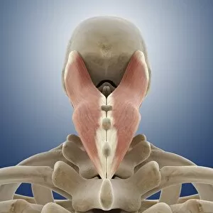
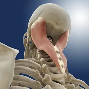
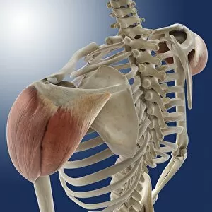
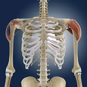
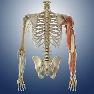
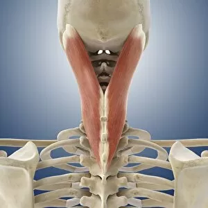
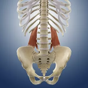
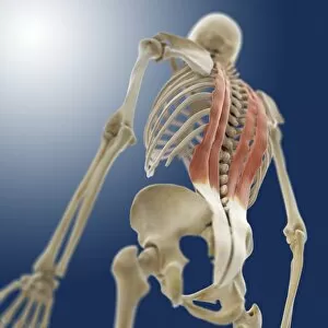
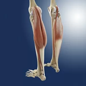
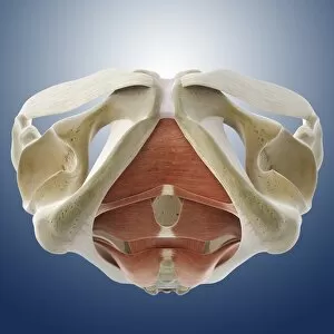
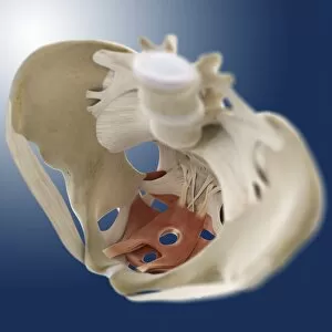
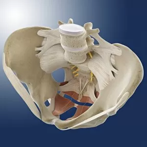
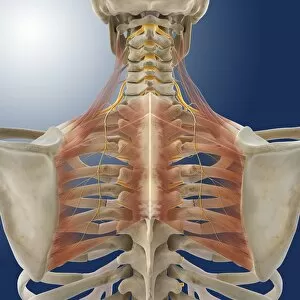
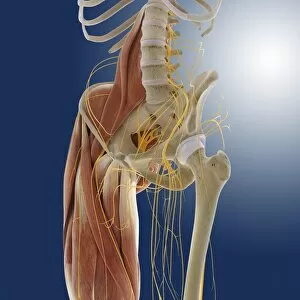
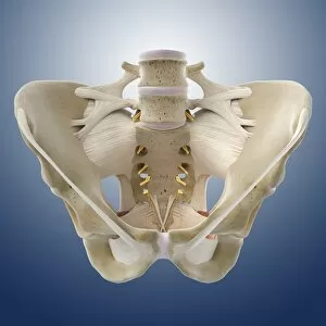
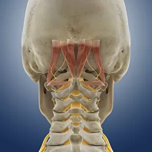
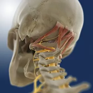
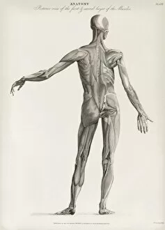
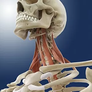
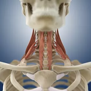
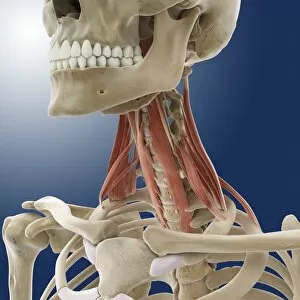
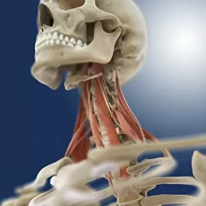
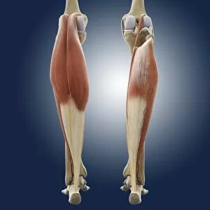
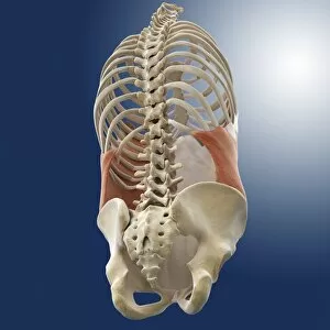
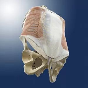
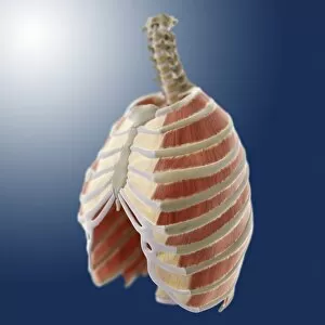
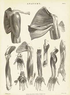
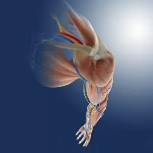
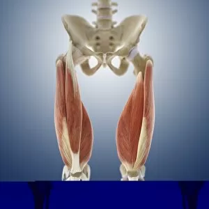
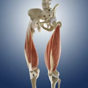
"The Marvels of the Muscular System: A Visual Journey through the Human Body" Discovering the intricate web of facial muscles that give us expressions - a fascinating study in human emotions. (Facial muscles of the human face with labels) Unveiling the hidden strength within our heads - explore the muscular system on head and witness its incredible complexity. Behold, an artistic masterpiece showcasing male muscles in all their glory, capturing raw power and grace. (Male muscles, artwork) Julien Bougle's vibrant colored plates superimposed on the human body reveal a mesmerizing tapestry of interconnected muscles - a true work of art. (The human body with superimposed colored plates by Julien Bougle) Delicate yet resilient, outer ankle ligaments depicted as captivating artwork C013 / 4452 will leave you in awe of their role in maintaining stability. Dive into the intricacies of inner ankle ligaments portrayed as stunning artwork C013 / 4451 - witness how they support every step we take. Celebrating female strength and beauty through an exquisite portrayal of the female muscular system - a testament to empowerment. (Female muscular system, artwork) Explore an illustration that unveils every contour and curve, highlighting both elegance and resilience within the female muscular system. The anatomy behind those enviable gluteal muscles revealed – marvel at their importance for posture, movement, and overall fitness. (Anatomy of gluteal muscles in human buttocks) Graceful movements come alive through this enchanting illustration featuring a talented female dancer – showcasing her mastery over her own muscular symphony. Andreas Vesalius' oil painting captures his profound understanding of anatomy; delve into his world where science meets artistry. (Andreas Vesalius oil on canvas)