
Myofibril Collection
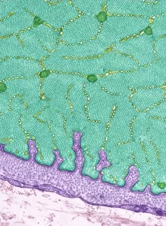
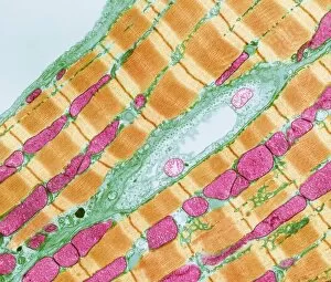
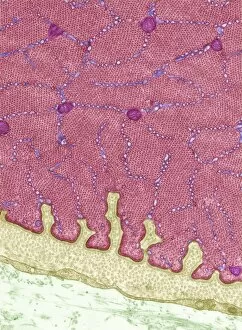
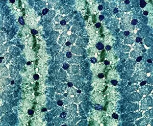
"Myofibril: The Mighty Machinery of Muscles Unveiled" Eye muscle, TEM C014 / 1468: Behold the intricate beauty of the myofibrils within our eye muscles


For sale as Licensed Images
Choose your image, Select your licence and Download the media
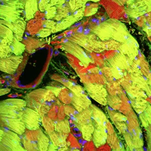
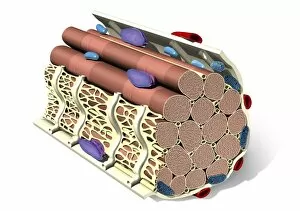
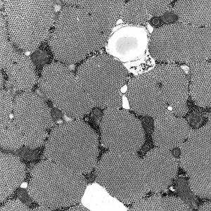
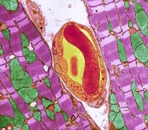
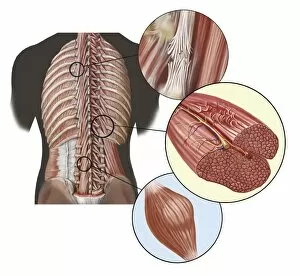
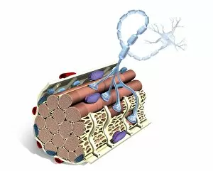
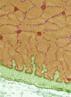
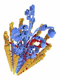
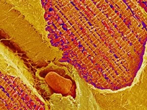
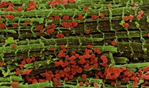
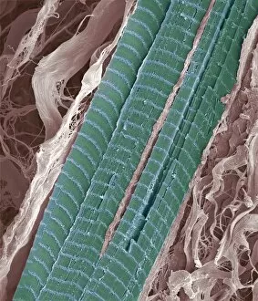
"Myofibril: The Mighty Machinery of Muscles Unveiled" Eye muscle, TEM C014 / 1468: Behold the intricate beauty of the myofibrils within our eye muscles, enabling us to see the world with clarity and precision. Heart muscle, confocal light micrograph: In this mesmerizing image, we witness the organized arrangement of myofibrils in cardiac muscle cells, ensuring our hearts beat rhythmically and tirelessly. Muscle fibre structure, artwork: A stunning artistic representation unveils the complex architecture of muscle fibers intertwined with myofibrils - a testament to their strength and resilience. Cardiac muscle, TEM: Underneath a microscope's lens lies an awe-inspiring view of cardiac muscle cells' myofibrillar network that powers every heartbeat with unwavering determination. Cardiac muscle and capillary, TEM: Witness how myofibrils intertwine seamlessly with tiny blood vessels in cardiac tissue - a harmonious collaboration essential for maintaining heart health. Detail of deep back muscles with a close-up of sprain, strain and spasm: Zooming into deep back muscles reveals the aftermaths of sprains, strains, and spasms – reminders that even mighty myofibrils need proper care to avoid injury. Torn muscle fibers with healing stages surrounding: Observe torn muscle fibers surrounded by healing processes as nature's remarkable ability repairs damaged tissues through coordinated efforts involving resilient myofibrils. Diagram of muscle structure: Unlocking the secrets within muscles' core is made easier through an informative diagram illustrating how various components including myofibrils work together flawlessly. Sugar uptake in muscles, diagram: Delve into sugar metabolism within active muscles as depicted in an enlightening diagram showcasing how energy is harnessed by these hardworking bundles called myofibrils. Actin Myosin Muscle Model, artwork C014 / 2661.