
Myoma Collection
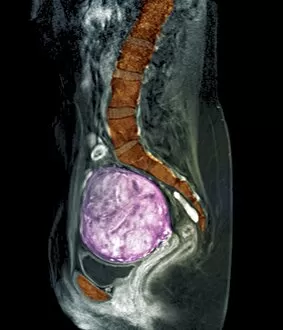
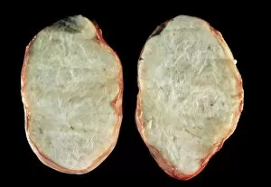
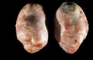
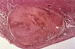
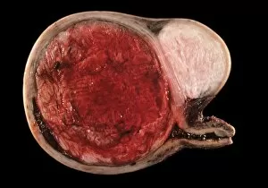
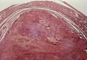
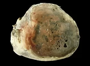
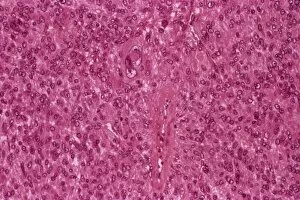
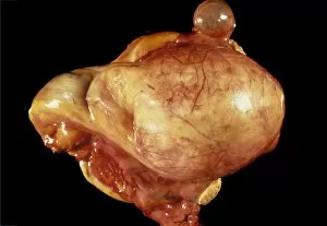
Myoma, also known as uterine fibroids, are non-cancerous growths that develop in the uterus


For sale as Licensed Images
Choose your image, Select your licence and Download the media
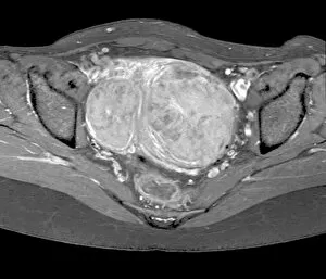
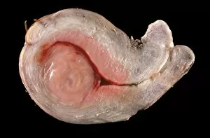
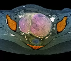
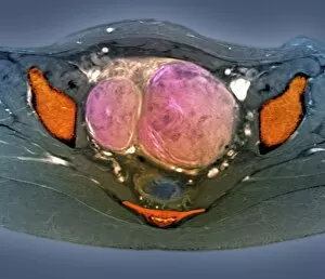
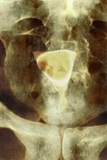
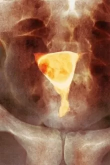
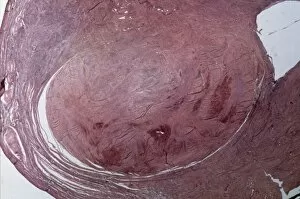
Myoma, also known as uterine fibroids, are non-cancerous growths that develop in the uterus. These abnormal growths can vary in size and number, causing a range of symptoms such as heavy menstrual bleeding, pelvic pain, and frequent urination. To diagnose myoma accurately, medical professionals often rely on advanced imaging techniques like MRI scans and X-rays. The MRI scans with codes C018 / 0466 to C018 / 0469 provide detailed images of uterine fibroids from different angles, aiding doctors in determining their location and size. Similarly, X-rays with codes C014 / 4918 and C014 / 4919 offer insights into the presence of uterine fibroids by capturing images using radiation. This diagnostic tool helps identify any calcifications or abnormalities associated with these growths. For a closer look at myoma under a microscope, light micrographs are utilized. Images labeled C015 / 7101 showcase a microscopic view of uterine fibroids while those marked as C015 / 6749 and C015 / 6748 depict ovarian fibroids. Additionally, light micrographs coded as C015/6737 and C015/6413 highlight the intricate details of uterine fibroid tissue structures. Understanding myoma through various imaging methods is crucial for accurate diagnosis and treatment planning. Whether it's an MRI scan or an X-ray or even examining samples under a microscope – each technique contributes to providing comprehensive information about this common condition affecting many women worldwide (Uterine Fibroid: Light Micrograph -C015/6416). By utilizing these tools effectively, healthcare professionals can tailor appropriate treatment options for patients dealing with myoma.