
Narrowed Collection (#2)
"Narrowed: Unveiling the Intricacies of Cardiovascular Health" In a captivating journey through medical imagery, we explore the world arteries and their treatments


For sale as Licensed Images
Choose your image, Select your licence and Download the media
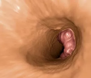
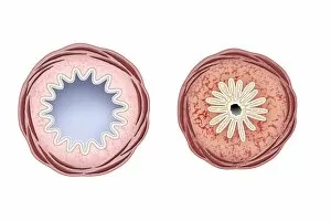
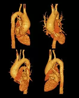
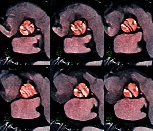
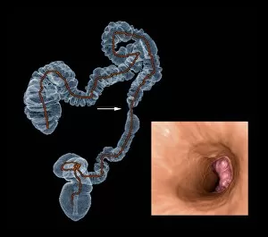
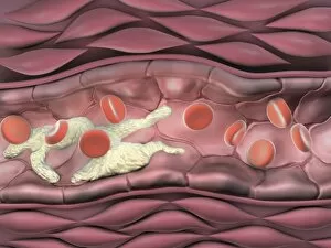
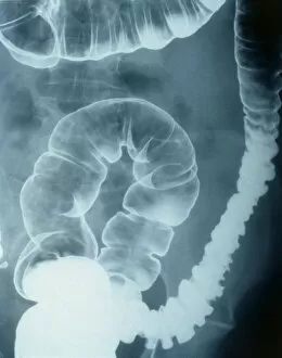
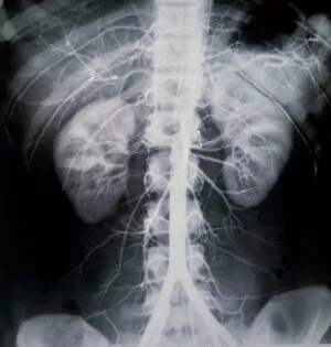
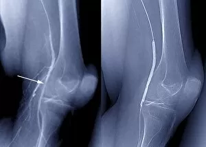
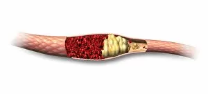
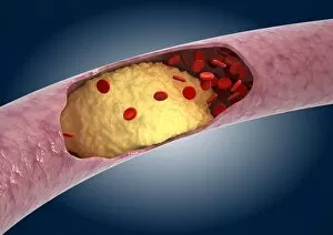
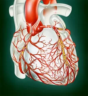
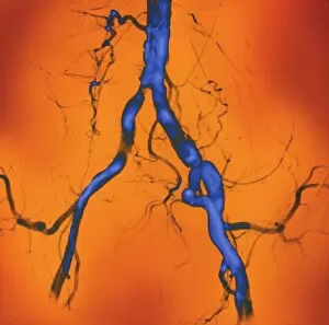
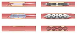
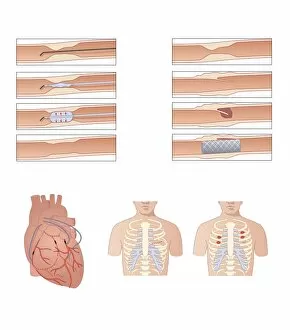
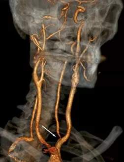
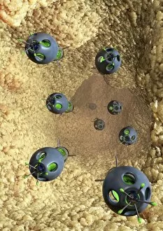
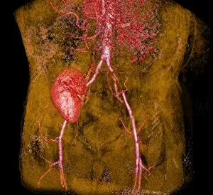
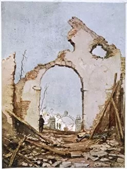
"Narrowed: Unveiling the Intricacies of Cardiovascular Health" In a captivating journey through medical imagery, we explore the world arteries and their treatments. Curtis British Entomology Plate 542 serves as an unexpected parallel to our own intricate vascular system, reminding us that even nature's smallest creations can hold profound lessons. Balloon angioplasty, depicted in X-ray form, emerges as a beacon of hope for those battling blocked arteries. The delicate procedure delicately widens constricted pathways, allowing life-giving blood to flow freely once more. Computer artwork vividly illustrates the complexity involved in this groundbreaking technique. Aortic dissection comes into focus with a stunning 3D CT scan, revealing the dangerous consequences of weakened arterial walls. As we witness this condition unfold before our eyes, it becomes evident why early detection is crucial in preventing catastrophic events. The silent menace known as atherosclerosis takes center stage through evocative artwork. This stealthy disease silently narrows arteries over time, gradually compromising cardiovascular health. Through these visual representations, we are reminded of the importance of adopting healthy lifestyles to thwart its progression. X-rays capture both pre and post-treatment stages of coronary stenosis – a condition where narrowing occurs within vital heart vessels. These powerful images showcase how medical interventions can restore vitality and improve quality of life for patients facing such challenges. As we delve deeper into these X-ray snapshots documenting coronary stenosis treatment journeys, each image tells its own story – one filled with resilience and triumph over adversity. Witnessing arteries transform from obstructed passageways to restored conduits reinforces the remarkable capabilities of modern medicine. Ultimately, these mesmerizing visuals serve as reminders that even when faced with narrow paths or compromised health conditions like coronary stenosis or atherosclerosis.