Nerves Collection (#5)

"Nerves: Unraveling the Intricacies of our Body's Communication System" Our bodies are a complex network of interconnected systems

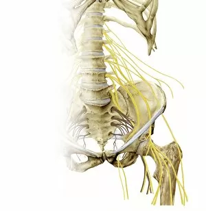
Background imageNerves Collection: Left hip and nerve plexus, artwork C016 / 6808

Left hip and nerve plexus, artwork C016 / 6808
Left hip and nerve plexus. Artwork of the nerves (yellow) and bones of the left hip, seen from the front. This is the lumbar plexus, itself forming part of the lumbosacral plexus
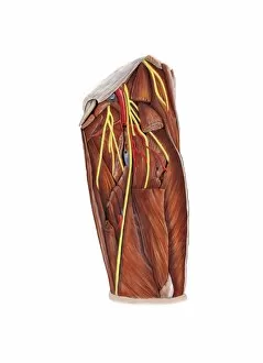
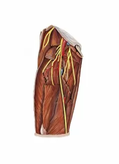
Background imageNerves Collection: Thigh nerve plexus, artwork C016 / 6804

Thigh nerve plexus, artwork C016 / 6804
Thigh nerve plexus. Artwork of the nerves (yellow), arteries (red), veins (blue), and muscles (brown) of the upper leg (thigh)
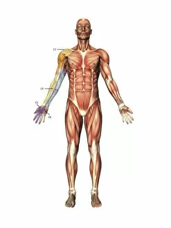
Background imageNerves Collection: Arm nerve regions, artwork C016 / 6840

Arm nerve regions, artwork C016 / 6840
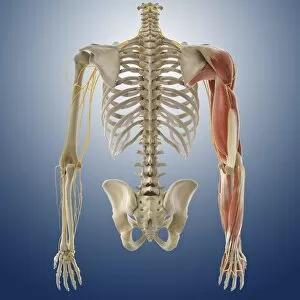
Arm nerve regions. Artwork of the muscles of the human body from the front, with the right arm (left) labelled with the names of the spinal nerves (cervical, neck, C; and thoracic, chest)

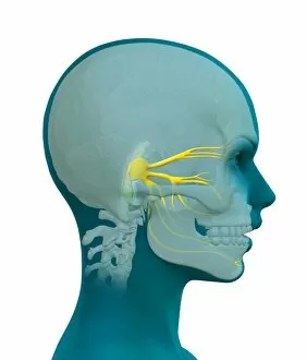
Background imageNerves Collection: Trigeminal nerve, artwork C016 / 7701

Trigeminal nerve, artwork C016 / 7701
Trigeminal nerve. Computer artwork showing the location of the trigeminal nerve (yellow) in a human head

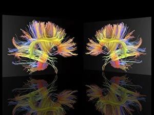
Background imageNerves Collection: White matter fibres of the human brain C016 / 9017

White matter fibres of the human brain C016 / 9017
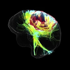
White matter fibres. Coloured 3D diffusion spectral imaging (DSI) scan of the bundles of white matter nerve fibres in the brain, depicting mirror neurons. Mirror neurons are found in the brain

Background imageNerves Collection: Right hip and nerve plexus, artwork C016 / 6809

Right hip and nerve plexus, artwork C016 / 6809
Right hip and nerve plexus. Artwork of the nerves (yellow) and bones of the right hip, seen from the front. This is the lumbar plexus, itself forming part of the lumbosacral plexus

Background imageNerves Collection: Right shoulder nerve plexus, artwork C016 / 6815

Right shoulder nerve plexus, artwork C016 / 6815
Right shoulder nerve plexus. Artwork of the nerves (yellow), arteries (red), muscles (red-brown) and bones (light brown) of the right shoulder, seen from the front

Background imageNerves Collection: Knee nerve plexus, artwork C016 / 6806

Knee nerve plexus, artwork C016 / 6806
Knee nerve plexus. Artwork of the nerves (yellow), arteries (red), veins (blue), and muscles (brown) of the rear of the knee

Background imageNerves Collection: Thigh nerve plexus, artwork C016 / 6805

Thigh nerve plexus, artwork C016 / 6805
Thigh nerve plexus. Artwork of the nerves (yellow), arteries (red), veins (blue), and muscles (brown) of the upper leg (thigh)

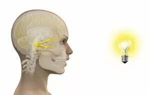
Background imageNerves Collection: Photic sneeze reflex, artwork C016 / 7700

Photic sneeze reflex, artwork C016 / 7700
Photic sneeze reflex. Computer artwork representing one explanation for the photic sneeze reflex. About a quarter of the population sneeze when they look at a bright light

Background imageNerves Collection: Left armpit nerve plexus, artwork C016 / 6812

Left armpit nerve plexus, artwork C016 / 6812
Left armpit nerve plexus. Artwork of the nerves (yellow), arteries (red), muscles (red-brown) and bones (light brown) of the left armpit, seen from the front

Background imageNerves Collection: Knee nerve plexus, artwork C016 / 6807

Knee nerve plexus, artwork C016 / 6807
Knee nerve plexus. Artwork of the nerves (yellow), arteries (red), veins (blue), and muscles (brown) of the rear of the knee

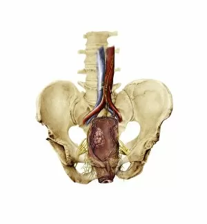
Background imageNerves Collection: Total mesorectal excision, artwork C016 / 6996

Total mesorectal excision, artwork C016 / 6996
Total mesorectal excision (TME). Artwork of the pelvis and the area of the colon and rectum that is removed in total mesorectal excision

Background imageNerves Collection: Synapse, artwork C016 / 7535

Synapse, artwork C016 / 7535
Synapse. Computer artwork of a synapse, the junction between nerve cells (orange). Synapses transmit electrical signals from one nerve cell to the next

Background imageNerves Collection: Left shoulder and nerve plexus, artwork C016 / 6810

Left shoulder and nerve plexus, artwork C016 / 6810
Left shoulder and nerve plexus. Artwork of the nerves (yellow), arteries (red), and bones of the left shoulder, seen from the front

Background imageNerves Collection: Left shoulder nerve plexus, artwork C016 / 6814

Left shoulder nerve plexus, artwork C016 / 6814
Left shoulder nerve plexus. Artwork of the nerves (yellow), arteries (red), muscles (red-brown) and bones (light brown) of the left shoulder, seen from the front

Background imageNerves Collection: White matter fibres of the human brain C016 / 9012

White matter fibres of the human brain C016 / 9012
White matter fibres. Coloured 3D diffusion spectral imaging (DSI) scan of the bundles of white matter nerve fibres in the brain, depicting mirror neurons. Mirror neurons are found in the brain

Background imageNerves Collection: Right armpit nerve plexus, artwork C016 / 6813

Right armpit nerve plexus, artwork C016 / 6813
Right armpit nerve plexus. Artwork of the nerves (yellow), arteries (red), muscles (red-brown) and bones (light brown) of the right armpit, seen from the front

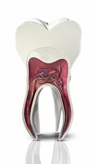
Background imageNerves Collection: Tooth cross-section, artwork

Tooth cross-section, artwork. The upper (biting) surfaces of the tooth are at top, with the lower sections (bottom) embedded in the gums and jaw bone (not shown)

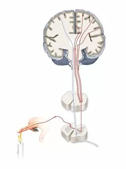
Background imageNerves Collection: Pain pathways, artwork

Pain pathways, artwork
Pain pathways, computer artwork. The heat of the candle is detected by nociceptors (special sensory nerve endings) in the finger

Background imageNerves Collection: Nerve growth factor bound to receptor

Nerve growth factor bound to receptor, molecular model. Nerve growth factor (NGF) complexed with the TrkA receptor. NGF is a neurotrophin that acts on the development and function of nerves

Background imageNerves Collection: Myenteric nerve plexus, TEM

Myenteric nerve plexus, TEM
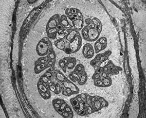
Myenteric nerve plexus. Transmission electron micrograph (TEM) of a section through a bundle of unmyelinated nerve fibres in the myenteric nerve plexus of the intestinal tract

Background imageNerves Collection: Smooth muscle of gut wall, TEM

Smooth muscle of gut wall, TEM
Smooth muscle of gut wall. Transmission electron micrograph (TEM) of a section through the outermost wall of the intestine

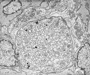
Background imageNerves Collection: Unmyelinated nerve, TEM

Unmyelinated nerve, TEM
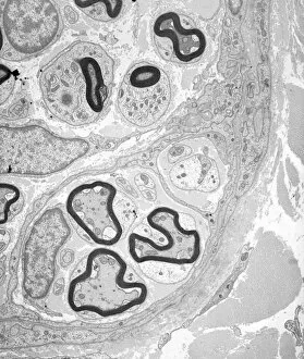
Unmyelinated nerve. Transmission electron micrograph (TEM) of a section through axon (nerve fibre) bundles of unmyelinated nerves

Background imageNerves Collection: Creutzfeldt-Jakob disease, MRI scan

Creutzfeldt-Jakob disease, MRI scan
Creutzfeldt-Jakob disease. Coloured magnetic resonance imaging (MRI) scan of an axial section through the brain of a 35-year-old patient

Background imageNerves Collection: Myelinated nerve tissue, TEM

Myelinated nerve tissue, TEM
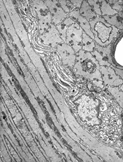
Myelinated nerve tissue. Transmission electron micrograph (TEM) of a section through myelinated nerve fibres (axons). Each axon is coated with many layers of myelin

Background imageNerves Collection: Olfactory bulb anatomy, artwork

Olfactory bulb anatomy, artwork
Olfactory bulb anatomy. Artwork of a human nose, showing the anatomy of the olfactory bulb, the organ of smell. This lies above the anterior aspect of the nasal cavity on the ethmoid bone

Background imageNerves Collection: Retina of the eye, light micrograph C016 / 0528

Retina of the eye, light micrograph C016 / 0528
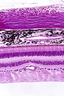
Retina of the eye. Light micrograph of a section through the retina from a human eye. From top down: nerve fibres of the optic nerve and a blood vessel; several layers of neurons (nerve cells)

Background imageNerves Collection: Nerve ganglion, light micrograph C016 / 0532

Nerve ganglion, light micrograph C016 / 0532
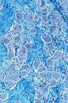
Nerve ganglion. Light micrograph of a section through a nerve ganglion of the peripheral nervous system, showing clusters of nerve cell bodies

Background imageNerves Collection: Myelinated nerve fibres, light micrograph C016 / 0531

Myelinated nerve fibres, light micrograph C016 / 0531
Myelinated nerve fibres. Light micrograph of a section through a spinal sensory nerve containing many closely packed axons exhibiting a wave-type appearance

Background imageNerves Collection: Retina layers, SEM C018 / 0521

Retina layers, SEM C018 / 0521
Retina layers, coloured scanning electron micrograph (SEM). The retina of the human eye is found on the inside of the eyeball and is formed from a number of layers

Background imageNerves Collection: Hand anatomy, illustration C018 / 0796

Hand anatomy, illustration C018 / 0796
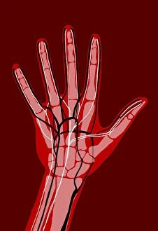
Hand anatomy. Illustration showing the bones, nerves and blood vessels of the hand. There are 27 bones, made up of 14 phalanges on the fingers and thumb

Background imageNerves Collection: Inner ear anatomy C018 / 6400

Inner ear anatomy C018 / 6400
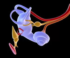
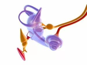
Inner ear anatomy. Computer artwork of a section through the inner part of a human ear, the organ of hearing and balance. The eardrum transmits sounds, as vibrations

Background imageNerves Collection: Corpus callosum, DTI MRI scan C017 / 7045

Corpus callosum, DTI MRI scan C017 / 7045
Corpus callosum. Coronal 3D diffusion tensor imaging (DTI) magnetic resonance imaging (MRI) scan of nerve pathways (blue) in and around the brains corpus callosum

Background imageNerves Collection: Inner ear anatomy C018 / 6379

Inner ear anatomy C018 / 6379
Inner ear anatomy. Computer artwork of a section through the inner part of a human ear, the organ of hearing and balance. The eardrum transmits sounds, as vibrations

Background imageNerves Collection: Inner ear anatomy C018 / 6393

Inner ear anatomy C018 / 6393
Inner ear anatomy. Computer artwork of a section through the inner part of a human ear, the organ of hearing and balance. The eardrum transmits sounds, as vibrations

Background imageNerves Collection: Inner ear anatomy C018 / 6387

Inner ear anatomy C018 / 6387
Inner ear anatomy. Computer artwork of a section through the inner part of a human ear, the organ of hearing and balance. The eardrum transmits sounds, as vibrations

Background imageNerves Collection: Inner ear anatomy C018 / 6397

Inner ear anatomy C018 / 6397
Inner ear anatomy. Computer artwork of a section through the inner part of a human ear, the organ of hearing and balance. The eardrum transmits sounds, as vibrations

Background imageNerves Collection: Inner ear anatomy C018 / 6402

Inner ear anatomy C018 / 6402
Inner ear anatomy. Computer artwork of a section through the inner part of a human ear, the organ of hearing and balance. The eardrum transmits sounds, as vibrations

Background imageNerves Collection: Female anatomy, artwork C017 / 0695

Female anatomy, artwork C017 / 0695
Female anatomy, artwork

Background imageNerves Collection: Bernhard von Gudden, German anatomist C018 / 7084

Bernhard von Gudden, German anatomist C018 / 7084
Bernhard von Gudden (1824-1886). 1885 engraving of the German microanatomist and psychiatrist Johann Bernhard Aloys von Gudden

Background imageNerves Collection: Glioblastoma brain tumour, DTI MRI scan C017 / 7052

Glioblastoma brain tumour, DTI MRI scan C017 / 7052
Glioblastoma brain tumour. 3D diffusion tensor imaging (DTI) magnetic resonance imaging (MRI) scan of nerve pathways (coloured) around a glioblastoma (upper right) in the brain

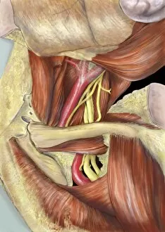
Background imageNerves Collection: Head and neck anatomy, artwork C014 / 0512

Head and neck anatomy, artwork C014 / 0512
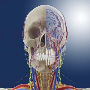
Head and neck anatomy, computer artwork. Arteries are red, veins are blue, nerves are deep yellow and lymph vessels are bright yellow

Background imageNerves Collection: Head and neck anatomy, artwork C014 / 0509

Head and neck anatomy, artwork C014 / 0509
Head and neck anatomy, computer artwork. The skin is transparent allowing arteries (red), veins (blue), nerves (deep yellow) and lymph vessels (bright yellow) to be seen

Background imageNerves Collection: Severe headache, artwork

Severe headache, artwork
Severe headache. Artwork of a man holding his head in pain. The glowing red area represents severe localised headaches such as cluster headaches, and can also represent migraines

Background imageNerves Collection: Human hand, historical anatomical model

Human hand, historical anatomical model
Human hand, anatomical model. 1950s model showing the muscle structure (red) and nerves (blue) of a human left hand

Background imageNerves Collection: Arm muscles, artwork

Arm muscles, artwork
Arm muscles, computer artwork. The uppermost muscle, wrapping around the shoulder, is the deltoid (dark green). Below it are the triceps

Background imageNerves Collection: Lumbar spinal nerves, 1825 artwork

Lumbar spinal nerves, 1825 artwork
Lumbar spinal nerves. Lateral view of the lower vertebral column, showing nerves (white) of the sympathetic nervous system. The pelvic ilium bone has been removed


For sale as Licensed Images

Choose your image, Select your licence and Download the media

"Nerves: Unraveling the Intricacies of our Body's Communication System" Our bodies are a complex network of interconnected systems, and at the heart of it all lies our nervous system. From chakras to brain fibers, this captivating web controls every sensation we experience. Delving deeper into the mysteries, DTI MRI scans like C017/7099 and C017/7035 reveal intricate brain pathways that transmit signals throughout our body. These scans provide us with a glimpse into the fascinating world within. A diagram showcasing the human brain and spinal column reminds us of the delicate balance maintained by these vital structures, and is through these pathways that information flows seamlessly, allowing us to perceive and react to our surroundings. Even in lighthearted moments, such as a dentist cartoon by H. M. Bateman depicting cause and effect, we can see how nerves play their part in triggering reactions – both physical and emotional. The inner ear is another marvel worth exploring; its auditory canal, eardrum, semicircular canals, cochlea nerve, and eustachian tube work together harmoniously to process sound waves. Throughout history, scientists have tirelessly studied the nervous system's intricacies - an 18th-century depiction serves as a testament to humanity's enduring curiosity about this remarkable mechanism. Yet amidst all its wonders lie certain afflictions - migraines remind us that even they are sometimes falter under strain. Understanding facial nerves becomes crucial when dealing with conditions affecting expressions or sensations on our face. So next time you find yourself asking "Are you nervous?", take a moment to appreciate just how incredible your own nerve system truly is – for it enables you to feel emotions deeply while simultaneously coordinating countless bodily functions without conscious effort. Intricate yet resilient; sensitive yet powerful – our they can an awe-inspiring reminder of nature's brilliance woven within each one of us.