
Neurite Collection

Neurite, the intricate web of nerve cells that powers our body and mind

Background imageNeurite Collection: Nerve cell, SEM

Nerve cell, SEM
Nerve cell. Coloured scanning electron micrograph (SEM) of a neuron (nerve cell). The cell body is the central structure with neurites (long and thin structures) radiating outwards from it

Background imageNeurite Collection: Microscopic view of a unipolar neuron

Microscopic view of a unipolar neuron. A unipolar neuron is a type of neuron in which only one protoplasmic process (neurite) extends from the cell body

Background imageNeurite Collection: Anatomy structure of neurons

Anatomy structure of neurons

Background imageNeurite Collection: Schematic of the hypothalamus receiving nerve impulses from the body

Schematic of the hypothalamus receiving nerve impulses from the body and sending messages to the circulatory and nervous system

Background imageNeurite Collection: Nerve with myelin sheath, seen in lower right, connects with muscle

Nerve with myelin sheath, seen in lower right, connects with muscle. Blood vessel and immune cells are seen in the center and upper right of image

Background imageNeurite Collection: Cerebral cortex nerve cells C018 / 0062

Cerebral cortex nerve cells C018 / 0062
Cerebral cortex nerve cells. Light micrograph of a section through neurones (nerve cells, black) in the cerebral cortex of a human brain

Background imageNeurite Collection: Cerebral cortex nerve cells C013 / 9767

Cerebral cortex nerve cells C013 / 9767
Cerebral cortex nerve cells. Light micrograph of a section through neurones (nerve cells, black) in the cerebral cortex of a human brain

Background imageNeurite Collection: Nerve cell synapses, computer artwork

Nerve cell synapses, computer artwork
Nerve cell synapses. Computer artwork of the chemical synapses between two nerve cells, or neurons, (one red and one blue)

Background imageNeurite Collection: Synapse, computer artwork

Synapse, computer artwork
Synapses. Computer artwork of synapses, the junctions between the ends (blue, swollen) of two nerve cells (neurons). Nerve cells are responsible for passing information around the central nervous

Background imageNeurite Collection: Nerve cell growth

Nerve cell growth. Light micrograph of nerve cells (neurons) with immunofluorescent staining. These cells have been grown in culture

Background imageNeurite Collection: Nerve cell growth

Nerve cell growth. Fluorescent light micrograph of a PC12 cell following stimulation by nerve growth factor. The cell body contains the nucleus (green)

Background imageNeurite Collection: Nerve cell culture, SEM

Nerve cell culture, SEM
Nerve cell culture. Coloured scanning electron micrograph (SEM) of new growth from a cultured sample from a spinal cord. The numerous branching strands are neurites

Background imageNeurite Collection: Cerebral cortex nerve cells

Cerebral cortex nerve cells

Background imageNeurite Collection: Mirror neurons, conceptual image

Mirror neurons, conceptual image
Mirror neurons, conceptual computer artwork. Nerve cells, or neurons, and a human eye reflected on a shiny surface representing a mirror neuron cell. Mirror neurons are found in the brain

Background imageNeurite Collection: Mirror neuron, conceptual image

Mirror neuron, conceptual image
Mirror neuron, conceptual computer artwork. Nerve cell, or neuron, reflected in water, representing a mirror neuron cell. Mirror neurons are found in the brain

Background imageNeurite Collection: Neuromuscular junctions, artwork

Neuromuscular junctions, artwork
Neuromuscular junctions. Computer artwork of junctions between nerves (thread-like objects) and a muscle (purple surface)

Background imageNeurite Collection: Synapses, artwork

Synapses, artwork
Synapses. Computer artwork of synapses, the junctions between the ends (green, swollen) of two nerve cells (neurons). Signals are passed along nerve cells in the form of an electrical impulse


For sale as Licensed Images

Choose your image, Select your licence and Download the media

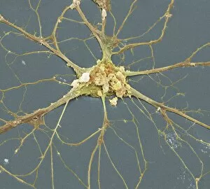
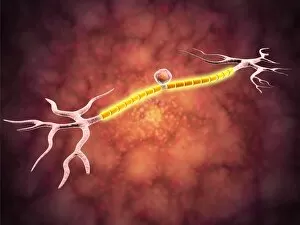
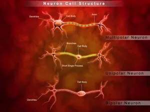
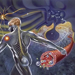
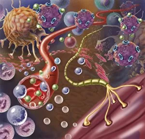
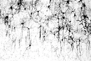
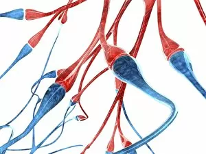
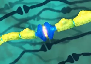
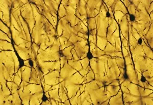
Neurite, the intricate web of nerve cells that powers our body and mind. 🧠✨ At a microscopic level, we can witness the mesmerizing beauty of neurites through scanning electron microscopy (SEM). These images reveal the delicate branches extending from nerve cells, forming an intricate network known as neural circuits. One captivating view showcases a unipolar neuron, displaying its unique structure in stunning detail. Neurites branch out from this central cell body, transmitting electrical signals throughout our nervous system. Zooming out to explore on a larger scale, we encounter the complex anatomy of neurons within our bodies. A schematic representation reveals how the hypothalamus receives crucial nerve impulses from various parts of our body, ensuring proper regulation and coordination. In another image, we observe a nerve with its protective myelin sheath connecting with muscle fibers. This essential connection allows for precise control over movement and coordination. Moving into the depths of our brain's cerebral cortex, we find ourselves surrounded by countless interconnected nerve cells. The images labeled C018 / 0062 and C013 / 9767 depict these fascinating structures responsible for higher cognitive functions such as memory and perception. Beyond biology's realm lies an intriguing parallel: artificial neural networks mimicking nature's design. Computer artwork portrays these simulated networks resembling real-life neurites – bridging science fiction with scientific reality. Whether it be in living organisms or computer simulations, neurites represent the foundation upon which information flows within neural networks, and are like highways carrying messages between different regions of our brains or facilitating communication in AI systems. So next time you marvel at your ability to think or move effortlessly - remember that it is thanks to these remarkable neurites orchestrating their symphony behind-the-scenes.