
Neurolemma Collection

Neurolemma, a crucial component of nerve fibers, plays a vital role in the myelination process

Background imageNeurolemma Collection: Myelination of nerve fibres, TEM

Myelination of nerve fibres, TEM
Myelination of nerve fibres. Coloured transmission electron micrograph (TEM) of Schwann cells (blue, with brown nuclei) insulating nerve fibres (axons, pink) with a myelin sheath

Background imageNeurolemma Collection: Myelination of nerve fibres, TEM

Myelination of nerve fibres, TEM
Myelination of nerve fibres. Coloured transmission electron micrograph (TEM) of Schwann cells (red, with blue nuclei) insulating nerve fibres (axons, orange) with a myelin sheath

Background imageNeurolemma Collection: Myelinated nerve, TEM

Myelinated nerve, TEM
Myelinated nerve. Coloured transmission electron micrograph (TEM) of myelinated nerve fibres and Schwann cells. Myelin (purple) is an insulating fatty layer that surrounds nerve fibres (axons)

Background imageNeurolemma Collection: Myelinated nerve, TEM C016 / 5840

Myelinated nerve, TEM C016 / 5840
Myelinated nerve. Coloured transmission electron micrograph (TEM) of a section through a myelinated nerve fibre and Schwann cell

Background imageNeurolemma Collection: Myelinated nerve, TEM C016 / 5839

Myelinated nerve, TEM C016 / 5839
Myelinated nerve. Coloured transmission electron micrograph (TEM) of a section through a myelinated nerve fibre and Schwann cell

Background imageNeurolemma Collection: Myelinated nerve, TEM C016 / 5838

Myelinated nerve, TEM C016 / 5838
Myelinated nerve. Coloured transmission electron micrograph (TEM) of a section through a myelinated nerve fibre and Schwann cell

Background imageNeurolemma Collection: Myelinated nerve, TEM C016 / 5448

Myelinated nerve, TEM C016 / 5448
Myelinated nerve. Transmission electron micrograph (TEM) of a section through a myelinated nerve fibre and Schwann cell. Myelin (black)

Background imageNeurolemma Collection: Myelinated nerve, TEM C016 / 5370

Myelinated nerve, TEM C016 / 5370
Myelinated nerve. Transmission electron micrograph (TEM) of a section through a myelinated nerve fibre and Schwann cell (centre)

Background imageNeurolemma Collection: Myelinated nerves, SEM C013 / 7142

Myelinated nerves, SEM C013 / 7142
Myelinated nerves. Coloured scanning electron micrograph (SEM) of a section through the sciatic nerve, showing the myelinated nerve fibres (axons)

Background imageNeurolemma Collection: Myelinated nerves, SEM C013 / 7141

Myelinated nerves, SEM C013 / 7141
Myelinated nerves. Coloured scanning electron micrograph (SEM) of a section through the sciatic nerve, showing the myelinated nerve fibres (axons)

Background imageNeurolemma Collection: Myelinated nerves, SEM C013 / 7138

Myelinated nerves, SEM C013 / 7138
Myelinated nerves. Coloured scanning electron micrograph (SEM) of a section through a myelinated nerve fibre (axon, beige, centre) from the sciatic nerve

Background imageNeurolemma Collection: Nerve demyelination, TEM

Nerve demyelination, TEM
Nerve demyelination. Coloured transmission electron micrograph (TEM) of a section through a Schwann cell and a nerve fibre, showing the early collapse of its myelin sheath

Background imageNeurolemma Collection: Myelinated nerves, SEM

Myelinated nerves, SEM
Myelinated nerves. Coloured scanning electron micrograph (SEM) of a section through myelinated nerve fibres and Schwann cells


For sale as Licensed Images

Choose your image, Select your licence and Download the media

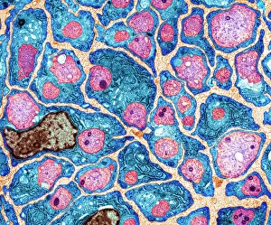
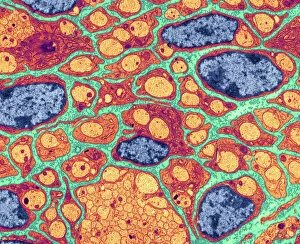
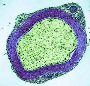
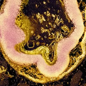
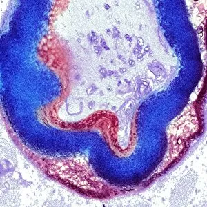
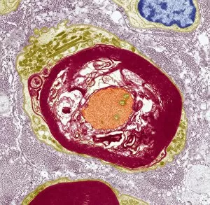
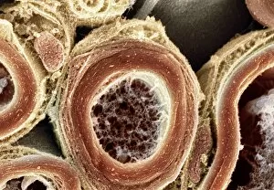
Neurolemma, a crucial component of nerve fibers, plays a vital role in the myelination process. Through transmission electron microscopy (TEM), scientists have been able to unravel the intricate details of myelinated nerves. The images captured through TEM reveal the mesmerizing beauty and complexity of these specialized cells. In one captivating image, we witness the myelination process in action. Nerve fibers are adorned with multiple layers of protective sheaths, forming a unique pattern that enhances their functionality. These layers not only provide insulation but also facilitate faster signal transmission along the nerve pathways. The TEM images showcase various stages of myelination, each more awe-inspiring than the last. From C016 / 5840 to C016 / 5370, we observe how these myelinated nerves undergo transformation and maturation. The intricate network formed by these fibers is truly remarkable. Furthermore, scanning electron microscopy (SEM) has allowed researchers to explore myelinated nerves from a different perspective. SEM images such as C013 / 7142 and C013 / 7141 offer an up-close view of this complex system. The detailed structures visible in these images highlight the precision with which nature has designed our neural networks. Studying neurolemma and its role in myelin formation provides valuable insights into neurological disorders like multiple sclerosis where demyelination occurs. By understanding how healthy nerve fibers function and maintain their integrity through proper myelin coverage, scientists can develop innovative therapies for those affected by such conditions. The world beneath our skin is filled with wonders waiting to be discovered; neurolemma's contribution to our nervous system is undoubtedly one of them. As research continues to unravel its mysteries using advanced imaging techniques like TEM and SEM, we inch closer towards unlocking new frontiers in neuroscience that could potentially revolutionize medical treatments for countless individuals worldwide.