Neurology Collection (#6)

Neurology, the fascinating study of the nervous system, unravels the intricate workings of our brain and its various components

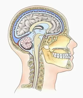
Background imageNeurology Collection: Lateral cross section through the skull showing the brain with intermediate layers (meninges)

Lateral cross section through the skull showing the brain with intermediate layers (meninges) highlighted

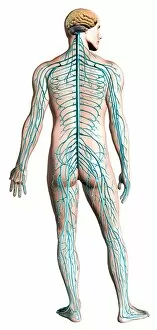
Background imageNeurology Collection: Diagram of human nervous system, posterior view

Diagram of human nervous system, posterior view

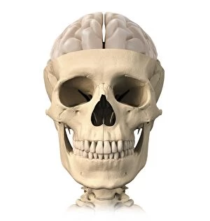
Background imageNeurology Collection: Anatomy of human skull, cutaway view with half brain showing

Anatomy of human skull, cutaway view with half brain showing, front view

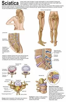
Background imageNeurology Collection: Medical chart showing the signs and symptoms of sciatica

Medical chart showing the signs and symptoms of sciatica

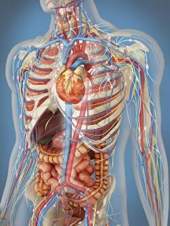
Background imageNeurology Collection: Human body showing heart and main circulatory system position

Human body showing heart and main circulatory system position
Transparent human body showing heart and main circulatory system position with internal organs, nervous system, lymphatic system and circulatory system

Background imageNeurology Collection: Human brain anatomy, superior view

Human brain anatomy, superior view

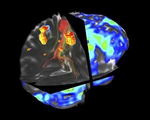
Background imageNeurology Collection: Brain tumour, fMRI and tractography C017 / 7102

Brain tumour, fMRI and tractography C017 / 7102
Brain tumour, fMRI and tractography. Combined functional magnetic resonance imaging (fMRI, blue and green) and tractography (yellow and red) imaging of a brain with a tumour (upper left)

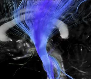
Background imageNeurology Collection: Corticospinal tract, DTI MRI scan C017 / 7046

Corticospinal tract, DTI MRI scan C017 / 7046
Corticospinal tract. Sagittal 3D diffusion tensor imaging (DTI) magnetic resonance imaging (MRI) scan of nerve pathways (blue) in the brains corticospinal tract

Background imageNeurology Collection: Normal human brain, MRI scan C016 / 8843

Normal human brain, MRI scan C016 / 8843
Brain. Coloured magnetic resonance imaging (MRI) scan of a sagittal section through a healthy human brain. The highly folded area (brown) is the cerebrum, which is responsible for conscious thought

Background imageNeurology Collection: Santiago Ramon y Cajal, histologist

Santiago Ramon y Cajal, histologist
Santiago Ramon y Cajal (1852-1934), Spanish histologist. Ramon y Cajal was apprenticed to a barber and a shoemaker before taking up medicine

Background imageNeurology Collection: Thomas Willis, 1621 - 1675. English neurologist, anatomist and author of the earliest known

Thomas Willis, 1621 - 1675. English neurologist, anatomist and author of the earliest known English work on medical psychology. Willis was a founding member of the Royal Society

Background imageNeurology Collection: Ivan Petrovich Pavlov, 1849 - 1936. Russian physiologist

Ivan Petrovich Pavlov, 1849 - 1936. Russian physiologist

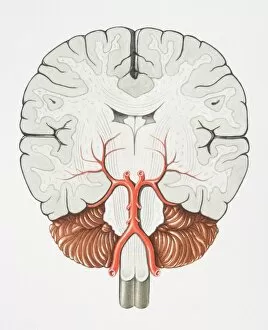
Background imageNeurology Collection: Coronal cross section of the Brain

Coronal cross section of the Brain

Background imageNeurology Collection: Human body showing autonomic nervous system and limbic system

Human body showing autonomic nervous system and limbic system
Side view of human body showing autonomic nervous system and limbic system within the brain. Green are parasympathetic nerves. Blue are sympathetic nerves
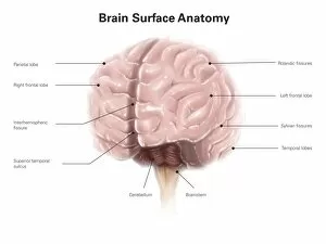
Background imageNeurology Collection: Brain surface anatomy, with labels

Brain surface anatomy, with labels

Background imageNeurology Collection: 3D rendering of human brain

3D rendering of human brain

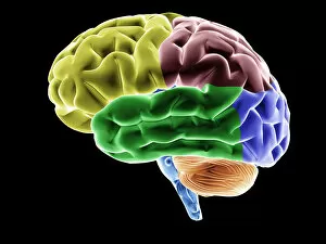
Background imageNeurology Collection: Side view of the human brain showing the functional lobes

Side view of the human brain showing the functional lobes

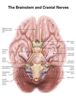
Background imageNeurology Collection: Anatomy of human brain stem and cranial nerves

Anatomy of human brain stem and cranial nerves

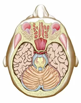
Background imageNeurology Collection: Transverse section of the midbrain

Transverse section of the midbrain

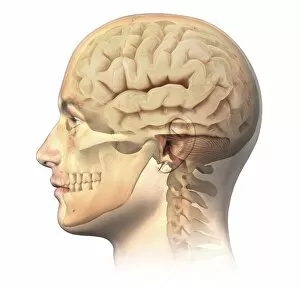
Background imageNeurology Collection: Anatomy of human head with skull and brain superimposed

Anatomy of human head with skull and brain superimposed, side view

Background imageNeurology Collection: Brain, various brain areas highlighted in colour, conceptual image for neurology, thinking

Brain, various brain areas highlighted in colour, conceptual image for neurology, thinking, knowledge, memory, anatomy, intelligence, 3D illustration

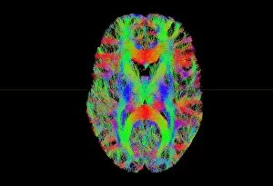
Background imageNeurology Collection: Brain fibres, tract density imaging C017 / 7039

Brain fibres, tract density imaging C017 / 7039
Brain fibres, tract density imaging. Axial scan of the brain showing the density of nerve pathways (tracts) in the brain. The image was obtained using 3D diffusion tensor imaging (DTI)
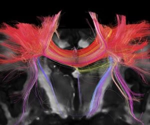
Background imageNeurology Collection: Corpus callosum, DTI MRI scan C017 / 7043

Corpus callosum, DTI MRI scan C017 / 7043
Corpus callosum. Coronal 3D diffusion tensor imaging (DTI) magnetic resonance imaging (MRI) scan of nerve pathways (red) in the brains corpus callosum

Background imageNeurology Collection: Brains white matter, DTI MRI scan

Brains white matter, DTI MRI scan
Brains white matter. Close-up of an area of the brain imaged using tract density imaging and 3D diffusion tensor imaging (DTI), a magnetic resonance imaging (MRI) technique

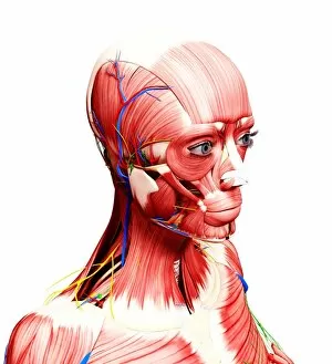
Background imageNeurology Collection: Female anatomy, artwork F007 / 3566

Female anatomy, artwork F007 / 3566
Female anatomy, computer artwork

Background imageNeurology Collection: Female anatomy, artwork F007 / 3972

Female anatomy, artwork F007 / 3972
Female anatomy, computer artwork

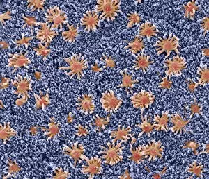
Background imageNeurology Collection: Nasal lining, SEM

Nasal lining, SEM
Nasal lining. Coloured scanning electron micrograph (SEM) of the olfactory epithelium that lines the nasal cavity, showing olfactory cells (red) surrounded by numerous cilia (hair-like projections)

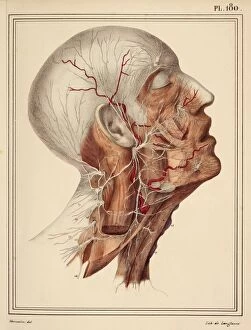
Background imageNeurology Collection: Face and neck nerves, 1825 artwork

Face and neck nerves, 1825 artwork
Face and neck nerves. Head and neck dissected to show the superficial nerves (white). The parotid gland (normally present below the ear)

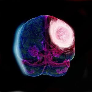
Background imageNeurology Collection: Brain tumour, DTI modelling C017 / 7060

Brain tumour, DTI modelling C017 / 7060
Brain tumour. Computer artwork of nerve pathways (coloured) in a brain with a tumour (white/red, upper right). The brain is seen from the front. Brain tumours can be benign or malignant (cancerous)

Background imageNeurology Collection: Sensory homunculus C017 / 6826

Sensory homunculus C017 / 6826
Model of a sensory homunculus. Parts of the body are sized according to how much space the brain gives to processing sensory information about that part of the body

Background imageNeurology Collection: Brain damage due to drugs, PET scans C014 / 1177

Brain damage due to drugs, PET scans C014 / 1177
Brain damage due to drugs, positron emission tomography (PET) scans. The brain at left is that of a control subject who had not taken drugs

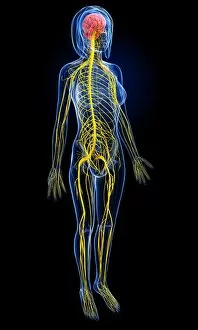
Background imageNeurology Collection: Female central nervous system, artwork

Female central nervous system, artwork
Female central nervous system, computer artwork

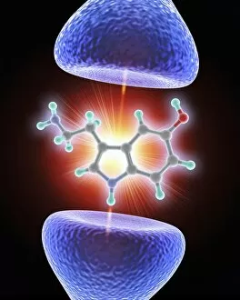
Background imageNeurology Collection: Nerve synapse and serotonin molecule

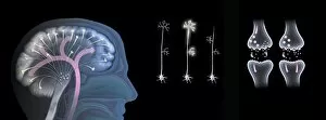
Nerve synapse and serotonin molecule
Nerve synapse and serotonin neurotransmitter molecule. Computer artwork of a junction, or synapse, between two nerve cells (neurons, blue)

Background imageNeurology Collection: Right shoulder and nerve plexus, artwork C016 / 6811

Right shoulder and nerve plexus, artwork C016 / 6811
Right shoulder and nerve plexus. Artwork of the nerves (yellow), arteries (red), and bones of the right shoulder, seen from the front

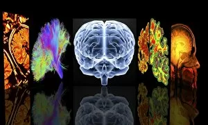
Background imageNeurology Collection: Neurology slide show C016 / 9008

Neurology slide show C016 / 9008
Conceptual computer artwork depicting neurology. From left to right: MRI brain scans, 3D dsi white matter brain scan, brain, Alzheimers brain versus normal brain, MRI brain scan

Background imageNeurology Collection: Brain blood supply, MRI scan C018 / 0432

Brain blood supply, MRI scan C018 / 0432
Brain haemorrhage. Coloured magnetic resonance imaging (MRI) scan of the brain of a 49 year old patient with a subdural haematoma (red)

Background imageNeurology Collection: Brain fibres, DTI MRI scan C017 / 7036

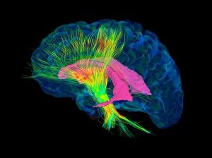
Brain fibres, DTI MRI scan C017 / 7036
Brain fibres. 3D diffusion tensor imaging (DTI) magnetic resonance imaging (MRI) scan of a selection of nerve pathways (green/yellow) in the brain

Background imageNeurology Collection: Valproic acid anticonvulsant molecule C014 / 2296

Valproic acid anticonvulsant molecule C014 / 2296
Valproic acid. Molecular model of the anticonvulsant and mood-stabilising drug valproic acid. It is used to treat epilepsy, seizures, bipolar disorder and depression

Background imageNeurology Collection: Effects of ecstasy on brain function

Effects of ecstasy on brain function. Computer artwork representing the effects of MDMA (3, 4-methylenedioxy-N-methylamphetamine), or ecstasy, on the nerves (centre) and synapses (nerve junctions)

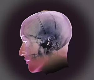
Background imageNeurology Collection: Deep brain stimulation, X-ray

Deep brain stimulation, X-ray
Image Xrays Crane (Sur Projection 3D Modèle) En Incidence De Profil Chez Une Patiente De 54 Ans, Parkinsonienne Traitee Sous Protocole De Neurochirurgie; Visualisation Des Electrodes De

Background imageNeurology Collection: Metropolis (1927) C016 / 4491

Metropolis (1927) C016 / 4491
Metropolis (1927). Poster for the the 1927 film Metropolis by Austrian film-maker Fritz Lang (1890-1976), showing the character Maria in Rotwangs transformation machine

Background imageNeurology Collection: Zebra fish embryo, SEM C013 / 9587

Zebra fish embryo, SEM C013 / 9587
Zebra fish embryo. Coloured scanning electron micrograph (SEM) of the embryo of a zebrafish (Danio rerio), showing its eyes either side of its large open mouth (centre)

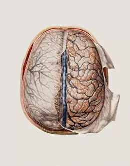
Background imageNeurology Collection: Brain meninges

Brain meninges, historical anatomical artwork. This cranial (top) view of the brain (with front at the top) and its surrounding protective meninges, which have been partially dissected away

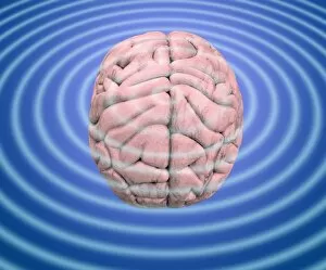
Background imageNeurology Collection: Brain waves, conceptual image

Brain waves, conceptual image
Brain waves. Conceptual image of waves emanating from a human brain. This can represent the electrical activity of the brain, as recorded on an electroencephalogram (EEG)

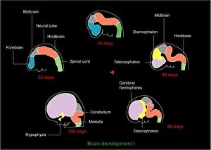
Background imageNeurology Collection: Foetal brain development, artwork

Foetal brain development, artwork. During the 4th week (upper left) the neural tube begins to differentiate into a spinal cord (green), forebrain (blue), midbrain (grey) and hindbrain (orange)

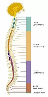
Background imageNeurology Collection: Spinal cord regions, diagram

Spinal cord regions, diagram
Spinal cord regions. Diagram of a sagittal section through the human skull and backbone, showing the five main regions of the spinal cord and the numbered nerves associated with them

Background imageNeurology Collection: Multiple sclerosis, MRI scan

Multiple sclerosis, MRI scan

Background imageNeurology Collection: Brain and spinal cord, artwork

Brain and spinal cord, artwork
Brain and spinal cord. Computer artwork showing the brain (top) and segments of the spinal cord. The spinal cord begins at the medulla oblongata at the base of the brainstem


For sale as Licensed Images

Choose your image, Select your licence and Download the media

Neurology, the fascinating study of the nervous system, unravels the intricate workings of our brain and its various components. From the Motor homunculus model that depicts how different body parts are represented in our motor cortex to the Histological Diagram of a Mammalian Retina revealing the complexity of our visual processing, every aspect is awe-inspiring. The Cerebellum tissue, as seen through a light micrograph, showcases its unique structure responsible for coordinating movement and balance. Meanwhile, an inferior view of the Anatomy of human brain highlights its remarkable organization and interconnectedness. Through Brain fibres captured by DTI MRI scans like C017 / 7099 or C017 / 7035, we gain insights into neural pathways that facilitate communication between different regions. These pathways form a complex network crucial for transmitting information throughout our brain. Exploring Brain blood vessels using a 3D angiogram (C007 / 1981) reveals their vital role in supplying oxygen and nutrients to sustain neuronal function. Additionally, understanding Motor and sensory homunculi helps us comprehend how specific areas within our brains control different bodily functions. Delving deeper into neurology brings us face-to-face with Medulla oblongata artwork depicting this critical region involved in regulating essential autonomic functions such as breathing and heart rate. Furthermore, observing Nerve and glial cells under a light microscope illustrates their diverse roles in supporting neurons' health and functionality. Finally, examining Synapse nerve junctions through TEM unveils these microscopic structures where electrical signals are transmitted from one neuron to another—a fundamental process underlying all brain activity. In essence, neurology takes us on an incredible journey deep within ourselves—unveiling mysteries at both macroscopic levels like anatomical structures or microscopically exploring cellular intricacies, and is through this exploration that we gain profound knowledge about how our brains work—the very essence of who we are as individuals.