
Neuropathology Collection
Neuropathology: Unveiling the Mysteries of the Brain In the realm of neuropathology, a fascinating world lies hidden beneath the surface of our minds

For sale as Licensed Images
Choose your image, Select your licence and Download the media
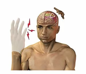
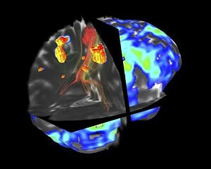
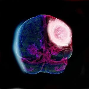
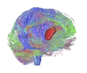
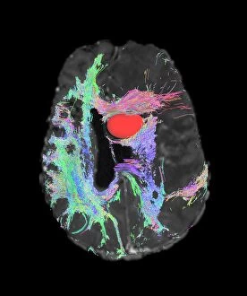
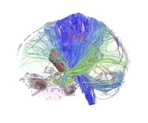
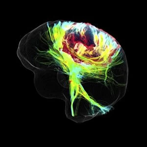
Neuropathology: Unveiling the Mysteries of the Brain In the realm of neuropathology, a fascinating world lies hidden beneath the surface of our minds, and is a domain where science and art converge to unravel the enigmatic workings of our brain. From phantom pain after amputation to intricate artwork depicting neural connections, this field delves into the complexities that make us who we are. One area that captivates researchers is studying brain tumors through advanced imaging techniques like fMRI and tractography. These cutting-edge methods, such as DTI modeling, allow scientists to visualize abnormalities in brain structure caused by glioblastoma tumors. The images captured during these scans, like C017 / 7102 or C017 / 7060, provide invaluable insights into understanding and treating these devastating conditions. Glioblastoma, an aggressive form of brain tumor, poses numerous challenges for both patients and doctors alike. Through DTI MRI scans (C017 / 7048-7059), experts can map out intricate fiber pathways within the brain affected by this malignant growth. This knowledge enables medical professionals to tailor treatment plans specifically targeting these areas while minimizing damage to healthy tissue. As we explore neuropathology further, it's essential to acknowledge pioneers like Edme Vulpian—a French neurologist whose contributions have shaped this discipline over time. His groundbreaking work paved the way for future generations in understanding neurological disorders and their underlying mechanisms. The study not only provides vital information on physical ailments but also sheds light on intangible aspects such as phantom pain experienced by amputees. By comprehending how our brains generate sensations from missing limbs (a phenomenon known as phantom limb syndrome), researchers can develop innovative therapies aimed at alleviating suffering and improving quality of life. Moreover, artistic expressions inspired by neuropathological findings offer a unique perspective on human cognition and emotions intertwined with scientific exploration.