
Nicolas Henri Jacob Collection
Nicolas Henri Jacob: A Master of Anatomy and Lithography Step into the fascinating world of Nicolas Henri Jacob


For sale as Licensed Images
Choose your image, Select your licence and Download the media
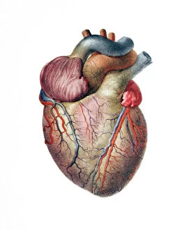
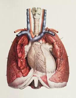
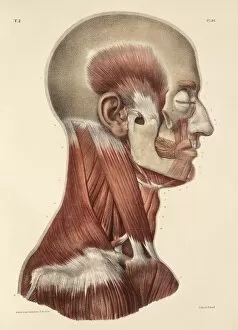
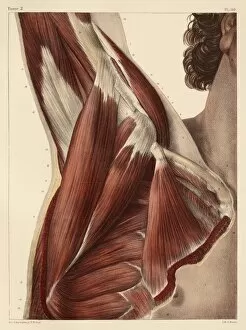
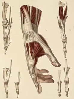
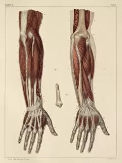
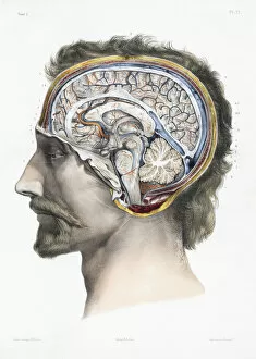
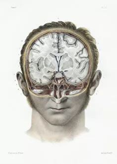
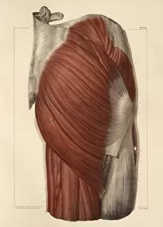
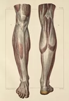
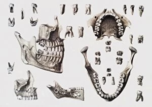
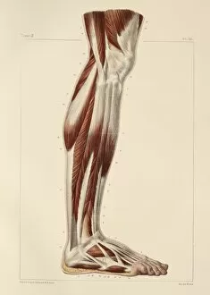
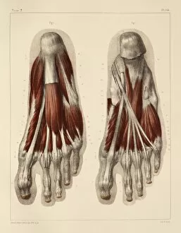
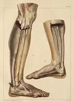
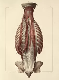
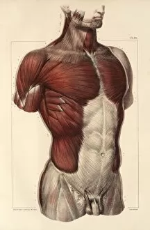
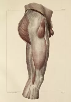
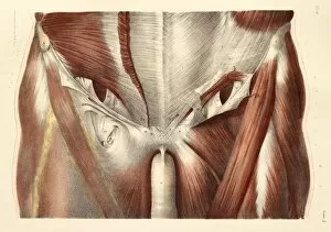
Nicolas Henri Jacob: A Master of Anatomy and Lithography Step into the fascinating world of Nicolas Henri Jacob, a French artist whose intricate artwork beautifully captures the intricacies of human anatomy. With his keen eye for detail and exceptional talent, Jacob brings to life the inner workings of our bodies in stunning illustrations. In his 1831 artworks, we witness the delicate intertwining of heart and brain blood vessels, showcasing their vital connection. The genius lies not only in his portrayal of these complex systems but also in how he effortlessly depicts the interplay between heart and lungs, ensuring our survival with every beat. Jacob's attention to detail extends beyond internal organs; he delves into head and neck muscles as well. His 1831 artwork reveals an exquisite understanding of facial expressions through intricate depictions of face and neck muscles, and is as if he has captured the essence of emotion within each stroke. One cannot overlook Jacob's contribution to lithography itself. In 1819, he created "The Genius of Lithography, " a testament to his mastery over this medium that revolutionized printing techniques during that era. Through this piece, we glimpse into both his artistic brilliance and technical expertise. His exploration continues with hand muscle anatomy from 1831—every tendon meticulously portrayed—a testament to his commitment to accuracy even in minute details. Similarly, muscles found in forearms are brought forth on paper with such precision that one can almost feel their strength emanating from the page. Venturing deeper into human physiology, Jacob collaborated with Jean-Baptiste Marc Bourgery on "Traite Complet de l'Anatomie de l'Homme. " Within its pages lie sagittal sections as well as vertical sections depicting various aspects of the human brain—an invaluable resource for medical knowledge during its time. As if unraveling secrets held within our bodies wasn't enough, Jacob turned his attention towards pelvic-femoral muscles—an area often overlooked.