
Nucleolus Collection (#2)
The nucleolus, a small but mighty organelle found within the nucleus of cells, plays a crucial role in various organisms


For sale as Licensed Images
Choose your image, Select your licence and Download the media
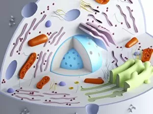
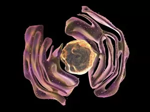
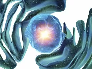
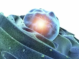
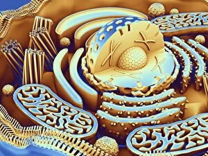
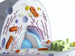
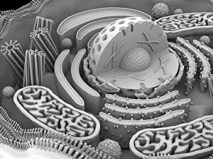
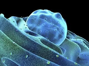
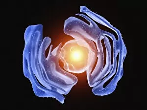
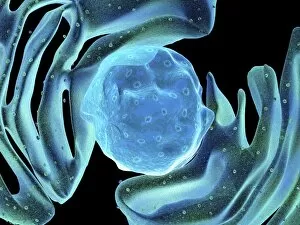
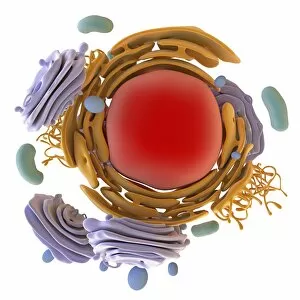
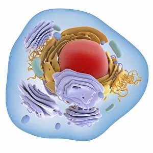
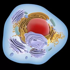
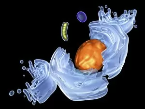
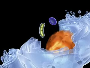
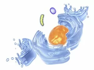
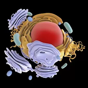
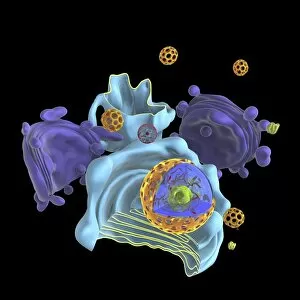
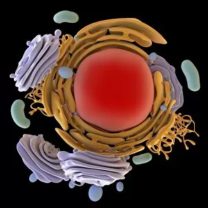
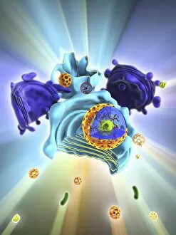
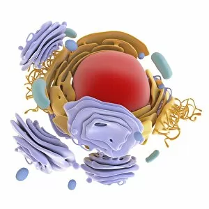
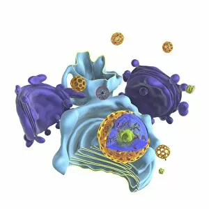
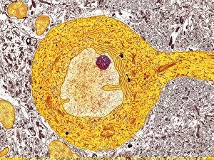
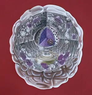
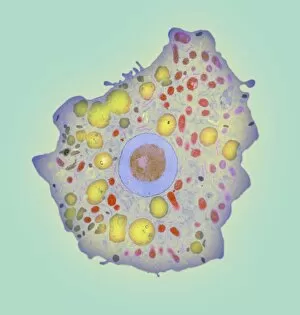
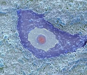
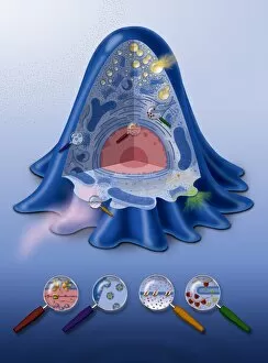
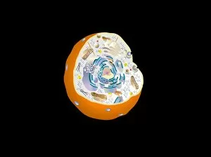
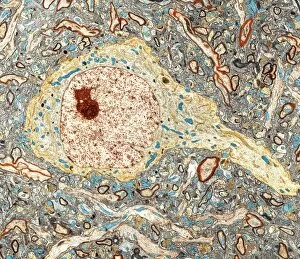
The nucleolus, a small but mighty organelle found within the nucleus of cells, plays a crucial role in various organisms. In budding yeast cells, it is responsible for ribosome biogenesis and assembly. This intricate process ensures that proteins are synthesized correctly to support cell growth and division. In Trypanosome protozoans, the nucleolus takes on a unique appearance as depicted in artwork. Its distinct structure aids in the production of ribosomal RNA (rRNA), which is essential for protein synthesis. When observing nerve cells under a transmission electron microscope (TEM), one can witness the presence of the nucleolus. These TEM images highlight its significance in these specialized cells involved in transmitting electrical signals throughout our bodies. Immunofluorescent light microscopy (LM) allows us to visualize fibroblast cell nuclei with their prominent nucleoli glowing brightly. This technique helps researchers study cellular processes such as DNA replication and gene expression. Purkinje nerve cells also exhibit an intriguing nucleolar structure when observed through TEM imaging techniques like C014 / 0583. These large neurons found in the cerebellum play a vital role in motor coordination. Artwork showcasing proteins, microtubules, and other cellular components emphasizes how interconnected they are with the nucleolus's functions. The accurate organization and functioning of these structures ensure proper cell division and overall cellular health. Cross-section illustrations provide insights into human cell composition where we find not only the nucleus but also its central component -the nucleolus- surrounded by other organelles like ribosomes and endoplasmic reticulum that work together harmoniously to maintain cell homeostasis. Plant cells possess similar nuclear structures including both nucleus and nucleolus along with additional features specific to plants such as chloroplasts or vacuoles necessary for photosynthesis or storage respectively; all working collectively towards plant growth and development.