Nucleus Collection (#21)

"The Nucleus: Unveiling the Mysteries of Life's Command Center" Exploring the intricate world within our cerebellum tissue

Background imageNucleus Collection: Vaccinia virus infected cell

Vaccinia virus infected cell. Immunofluorescence deconvolution micrograph of a cell infected with vaccinia virus particles. Host and viral DNA (deoxyribonucleic acid) is blue

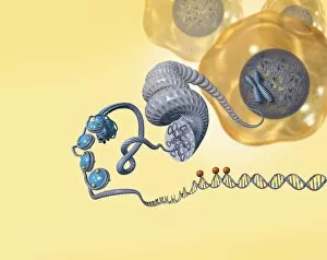
Background imageNucleus Collection: Cellular packaging of DNA, artwork

Cellular packaging of DNA, artwork
Cellular packaging of DNA. Artwork of a strand of the genetic material DNA (deoxyribonucleic acid) unwound from the nucleus (blue) of a cell (orange, upper right)

Background imageNucleus Collection: Fibroblast cells, fluorescent micrograph

Fibroblast cells, fluorescent micrograph
Fibroblast cells. Immunofluorescence deconvolution micrograph of fibroblast cells. The cell nuclei, which contain the cells genetic information, are blue

Background imageNucleus Collection: Nucleolus, SEM

Nucleolus, SEM
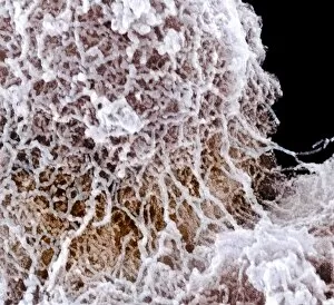
Nucleolus, coloured scanning electron micrograph (SEM). The nucleolus is responsible for producing components of ribosomes, the cells protein-manufacturing organelles

Background imageNucleus Collection: Plant cell, SEM

Plant cell, SEM
Plant cell. Coloured scanning electron micrograph (SEM) of a section through a plant cell, revealing its internal structure. The cell is encased in a cellulose, hemicellulose and pectin cell wall

Background imageNucleus Collection: Fat cells, TEM

Fat cells, TEM
Fat cells. Coloured transmission electron micrograph (TEM) of a section through fat (adipose) tissue. Each fat cell or adipocyte

Background imageNucleus Collection: Small lymphocyte, TEM

Small lymphocyte, TEM
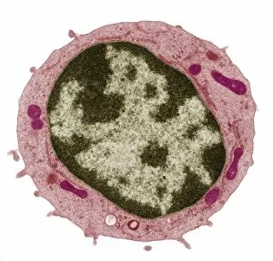
Small lymphocyte. Coloured transmission electron micrograph (TEM) showing the large central nucleus (brown) of a small lymphocyte (white blood cell)

Background imageNucleus Collection: White blood cell, TEM

White blood cell, TEM
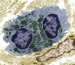
White blood cell. Coloured transmission electron micrograph (TEM) of an eosinophil (a type of white blood cell), part of the bodys immune system

Background imageNucleus Collection: Cell organelles, SEM

Cell organelles, SEM
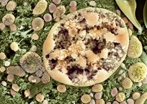
Cell organelles. Coloured scanning electron micrograph (SEM) of a section through a cell from a kidney proximal tubule. The cell nucleus (partially seen at top left)

Background imageNucleus Collection: Intestinal endocrine cell, TEM

Intestinal endocrine cell, TEM
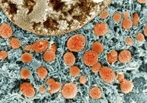
Intestinal endocrine cell, coloured transmission electron micrograph (TEM). This is a type 1 intestinal endocrine cell. The granules (red) contain serotonin

Background imageNucleus Collection: Neural stem cells in culture

Neural stem cells in culture
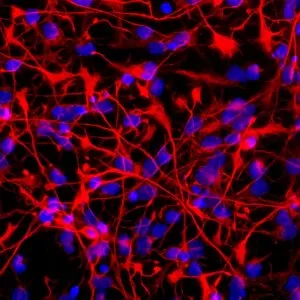
Neural stem cell in culture, fluorescent light micrograph. The stem cells have been dyed for nestin (red), an intermediate filament (IF) protein, and the nuclei are dyed blue

Background imageNucleus Collection: Brain cells in culture, light micrograph

Brain cells in culture, light micrograph
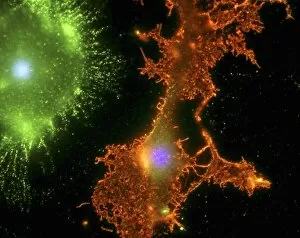
Brain cells in culture. Fluorescent light micrograph of a microglial cell (upper left) and an oligodendrocyte (centre) from a human brain

Background imageNucleus Collection: Cell nucleus, SEM

Cell nucleus, SEM
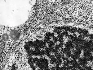
Cell nucleus. Coloured scanning electron micrograph (SEM) of a section through the nucleus (centre right) of a kidney cell

Background imageNucleus Collection: Cytoskeleton, confocal light micrograph

Cytoskeleton, confocal light micrograph. Tubulin, the protein that makes up microtubules, is blue. Microtubules are part of the cytoskeleton, which maintains the cells shape

Background imageNucleus Collection: Reptilian red blood cells, SEM

Reptilian red blood cells, SEM
Reptilian red blood cells. Coloured scanning electron micrograph (SEM) of whole and fractured red blood cells (erythrocytes, purple) from reptilian tissue

Background imageNucleus Collection: Nuclear envelope connecting to ER

Nuclear envelope connecting to ER
Transmission electron micrograph of section through part of a nucleus of a filament cell in moss Physcomitrella patens showing connection between nuclear envelope and endoplasmic reticulum

Background imageNucleus Collection: Chloroplasts in protoplast of tobacco

Chloroplasts in protoplast of tobacco
False-colour transmission electron micrograph of chloroplasts in a protoplast from a tobacco leaf, Nicotiana tabacum, (cultivar Xanthi)

Background imageNucleus Collection: Coloured TEM of a yeast cell

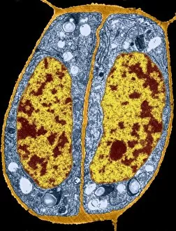
Coloured TEM of a yeast cell

Background imageNucleus Collection: Spirogyra algae, light micrograph

Spirogyra algae, light micrograph
Spirogyra algae. Light micrograph of filaments from a Spirogyra sp. alga, showing its spiralling bands of chloroplasts (green). Central nuclei (round) are also seen in each cell

Background imageNucleus Collection: Quark structure of silicon atom nucleus

Quark structure of silicon atom nucleus
Visualisation of a silicon nucleus. This image represents the nucleus of a silicon atom. The nucleus is made of 28 particles, called nucleons (14 protons and 14 neutrons)

Background imageNucleus Collection: High-energy cosmic ray collision

High-energy cosmic ray collision
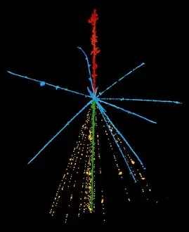
A very high-energy cosmic ray iron nucleus shoots into some photographic emulsion & collides with a silver or bromine nucleus in the emulsion to produce a tremendous " jet" of about 850

Background imageNucleus Collection: Emulsion photo of cosmic ray event

Emulsion photo of cosmic ray event
The power of a cosmic ray is shown in this false- colour emulsion photo of a cosmic ray sulphur nucleus (red) colliding with a nucleus in the emulsion

Background imageNucleus Collection: Visualisation of quark structure of uranium

Visualisation of quark structure of uranium
Quark structure of the uranium nucleus. Computer visualisation of the nucleus of a uranium atom. The most common isotope, uranium-238, consists of 92 protons and 146 neutrons
Background imageNucleus Collection: Art showing size of atomic components

Art showing size of atomic components
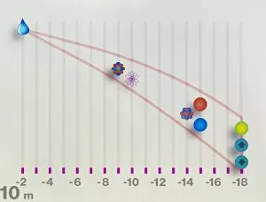
Atomic dimensions. Computer artwork showing the relative sizes of atoms and their components. The scale at bottom, measured in fractions of a metre, decreases from left to right

Background imageNucleus Collection: Stomatal complex, TEM

Stomatal complex, TEM
Stomal complex. Coloured transmission electron micrograph (TEM) of a stomatal (pore) complex in the young leaf of the pea plant (Pisum sativum)

Background imageNucleus Collection: Poppy ovary with developing seeds, LM

Poppy ovary with developing seeds, LM
Poppy ovary with developing seeds. Light micrograph (LM) of a cross-section through the centre of a mature poppy ovary, showing placentas bearing developing seeds (red)

Background imageNucleus Collection: Mature poppy ovary, light micrograph

Mature poppy ovary, light micrograph
Mature poppy ovary. Light micrograph (LM) of a cross-section through a mature poppy ovary, which is incompletely divided by septa (membranes) that do not meet in the centre

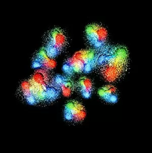
Background imageNucleus Collection: Visualisation of quark structure of gold

Visualisation of quark structure of gold

Background imageNucleus Collection: First photo of ultraheavy primary cosmic ray

First photo of ultraheavy primary cosmic ray
First picture of an ultraheavy cosmic ray primary, compared with a cosmic ray iron nucleus. The tracks of both nuclei are recorded in photographic emulsion exposed during balloon flights high in

Background imageNucleus Collection: Neon atom, artwork

Neon atom, artwork
Neon atom. Computer artwork of electron orbitals in a neon atom. The nucleus is represented by a flash of light. The orbitals shown are 1s (small white sphere)

Background imageNucleus Collection: Visualisation of quark structure of carbon

Visualisation of quark structure of carbon
Quark structure of the carbon nucleus. Computer visualisation of the nucleus of a carbon atom. The most common isotope, carbon-12, consists of six protons and six neutrons

Background imageNucleus Collection: High-energy cosmic ray

High-energy cosmic ray
A very high-energy cosmic ray iron nucleus shoots into some photographic emulsion & collides with a silver or bromine nucleus in the emulsion to produce a tremendous " jet" of about 850

Background imageNucleus Collection: Quark structure of carbon atom nucleus

Quark structure of carbon atom nucleus
Visualisation of a carbon nucleus. This image represents the nucleus of a carbon atom. The nucleus is made of twelve particles, called nucleons (six protons and six neutrons)


For sale as Licensed Images

Choose your image, Select your licence and Download the media

"The Nucleus: Unveiling the Mysteries of Life's Command Center" Exploring the intricate world within our cerebellum tissue, where the nucleus orchestrates every movement and balance. A mesmerizing light micrograph reveals the nucleus at the heart of M51 Whirlpool Galaxy, a cosmic dance guided by its gravitational pull. Witnessing life's beginning through a budding yeast cell, as its nucleus holds the blueprint for growth and reproduction. Delving into the secrets of communication with a nerve cell captured in stunning detail through SEM imaging, showcasing its complex nucleus. Behold the majestic Sombrero Galaxy (Messier 104), where billions of stars revolve around their central nucleus like celestial dancers in perfect harmony. The hippocampus brain tissue unveils memories etched within each neuron's nucleus, shaping who we are and what we remember. Peering into HeLa cells under a light microscope (C017/8299), revealing their remarkable nuclei that have revolutionized medical research. An artistic representation takes us deep into Medulla Oblongata's core—the vital center controlling essential bodily functions—where nuclei reign supreme. Captivating artwork depicts nuclear fission—a powerful force harnessed to generate energy while reminding us of its immense potential and responsibility. Marvel at NGC 4258 spiral galaxy's galactic light show, where vibrant colors illuminate star clusters revolving around their radiant nuclear core. Human cells come alive under scrutiny as their nuclei hold genetic information that shapes our unique characteristics and existence itself. Exploring kidney tubules in section unravels an intricate network governed by countless tiny nuclei working harmoniously to maintain our body’s equilibrium. Intricate yet awe-inspiring, these glimpses into various realms remind us that no matter how vast or minuscule, from galaxies to microscopic cells - all bow to the commanding presence of the nucleus.