
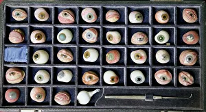
Ocular Collection
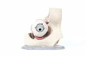
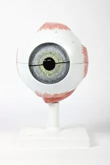
"Exploring the Intricate World Anatomy and Artwork" Step into a mesmerizing world where glass eyeballs come to life, showcasing the intricate beauty anatomy


For sale as Licensed Images
Choose your image, Select your licence and Download the media
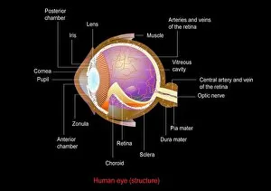
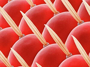
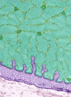
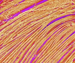
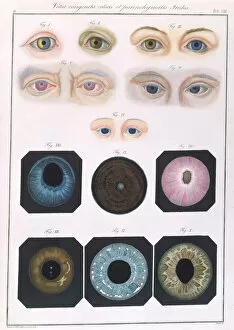
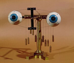
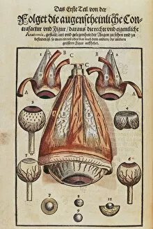
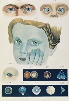
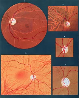
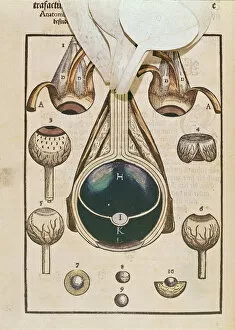
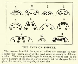
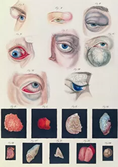
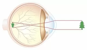
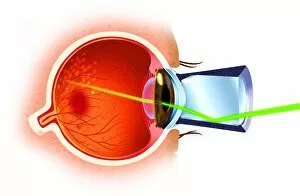
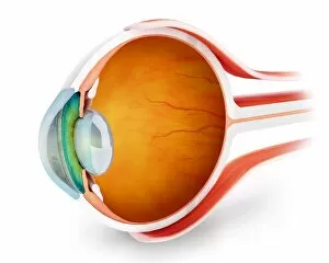
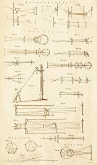
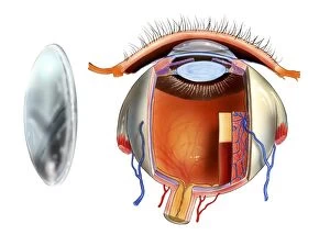
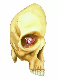
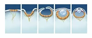
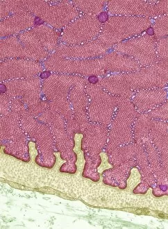
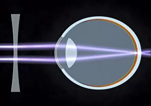
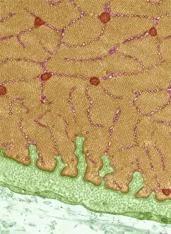
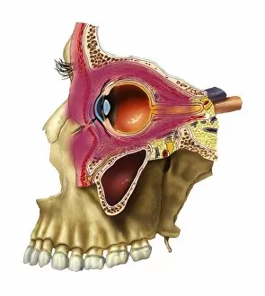
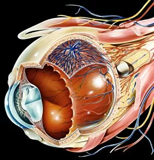
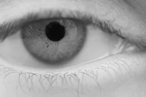
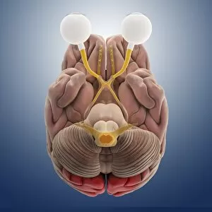
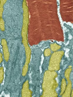
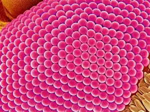
"Exploring the Intricate World Anatomy and Artwork" Step into a mesmerizing world where glass eyeballs come to life, showcasing the intricate beauty anatomy. From the delicate retina to the compound eye of a fly, every detail is captured in stunning SEM images like never before. Witness conceptual artwork depicting eye surgery, as skilled hands delicately manipulate this precious organ. Marvel at TEM images revealing the complexity of eye muscles, their fibers intricately woven together like an artistic masterpiece. Delve deeper into the inner workings of our eyes with SEM images that unveil the intricate network of blood vessels within the retina. These tiny highways carry life-sustaining nutrients and oxygen to ensure optimal vision. Explore further as SEM reveals detailed structures of eye muscles, highlighting their strength and flexibility in controlling our gaze. The magnified view showcases their fascinating composition and functionality. Travel back in time with Klinische Darstellungen der's description of congenital eye anomalies, offering insights into historical perspectives on ocular conditions. Witness how ophthalmological teaching apparatuses evolved over time through metal models designed for educational purposes. Finally, immerse yourself in Georg Bartisch's Ophthalmodouleia from centuries ago – a testament to early anatomical understanding. Discover his meticulous illustrations that laid foundations for modern-day knowledge about ocular anatomy. In this captivating journey through ocular artistry and scientific exploration, we gain a newfound appreciation for these remarkable organs that grant us sight – windows to both our external world and rich history.