
Ophthalmological Collection
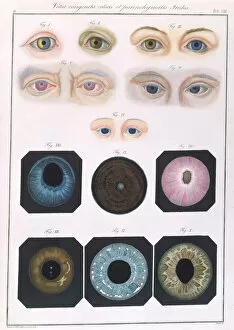
"Unveiling the Mysteries of Ophthalmological Anomalies


For sale as Licensed Images
Choose your image, Select your licence and Download the media
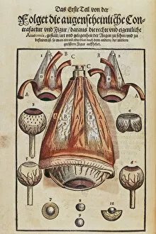
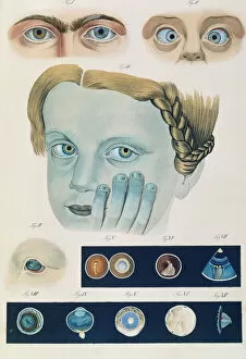
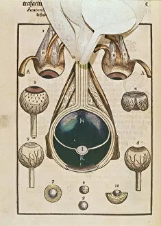
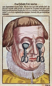
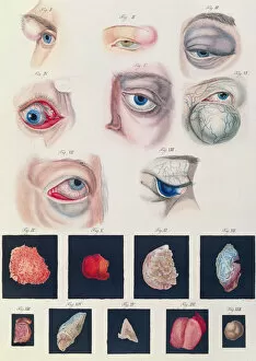
"Unveiling the Mysteries of Ophthalmological Anomalies: A Journey through Centuries" Step into the world of ophthalmology as we explore the captivating descriptions and illustrations of congenital eye anomalies from Klinische Darstellungen der. Delve deep into the intricate anatomy of the eye, as depicted in Georg Bartisch's Ophthalmodouleia, a timeless masterpiece from 1535-1636. Witness the remarkable mask used for treating strabismus, showcased in Ophthalmodouleia. Marvel at the innovative techniques employed to address various eye conditions, offering hope and relief to patients throughout history. Immerse yourself in an era where knowledge was scarce but determination prevailed. Discover how ancient physicians meticulously documented congenital eye anomalies, shedding light on these enigmatic conditions that have perplexed humanity for centuries. Join us on this enlightening journey as we unveil historical accounts of cataract operations and other groundbreaking procedures described in Ophthalmodouleia by Georg Bartisch (1535-1636). Witness firsthand how early surgeons courageously ventured into uncharted territories to restore vision and improve lives. Let us embark together on this odyssey through time, exploring both medical advancements and enduring mysteries within ophthalmology. From ancient texts to pioneering treatments, let our shared fascination with eyes guide us towards a brighter future for all who seek clarity and visual well-being.