Oral Collection (#3)

"Exploring the Intricacies of Oral Health

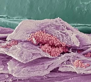
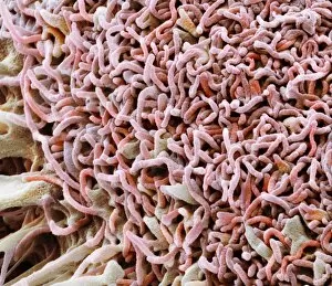
Background imageOral Collection: Thrush infection of the tongue, SEM C016 / 9091

Thrush infection of the tongue, SEM C016 / 9091
Thrush infection of the tongue. Coloured scanning electron micrograph (SEM) of the surface of a tongue infected with Candida sp. fungus (yellow), known as yeast infection, thrush, or candidiasis

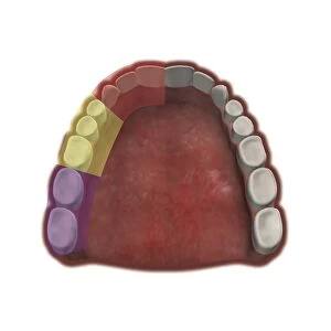
Background imageOral Collection: Dental maxillary nerve regions, artwork C016 / 6835

Dental maxillary nerve regions, artwork C016 / 6835
Dental maxillary nerve regions. Artwork of the teeth of the upper jaw (maxilla), seen from below, colour-coded by the branches of the superior alveolar nerve that supply the maxillary teeth

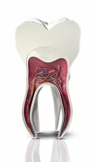
Background imageOral Collection: Tooth cross-section, artwork

Tooth cross-section, artwork. The upper (biting) surfaces of the tooth are at top, with the lower sections (bottom) embedded in the gums and jaw bone (not shown)

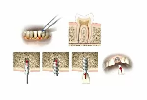
Background imageOral Collection: Dental implant, artwork

Dental implant, artwork
Dental implant. Artworks showing the process of replacing a tooth (incisor) with a dental implant. At upper left, front lower teeth and a dental instrument are shown in a case of receding

Background imageOral Collection: Light-reactive dental filling, artwork

Light-reactive dental filling, artwork
Light-reactive dental filling. Computer artwork showing a blue light (top left) being used to harden a composite dental filling in a tooth (lower right)

Background imageOral Collection: Root canal dental treatment, artwork

Root canal dental treatment, artwork
Root canal dental treatment. Computer artwork showing a drill (top left) being used to clear out the root canals (blue) of a decayed tooth

Background imageOral Collection: Mouth cancer cell, SEM C014 / 1460

Mouth cancer cell, SEM C014 / 1460
Mouth cancer cell. Coloured scanning electron micrograph (SEM) of a squamous cell carcinoma (cancer) cell from a human mouth

Background imageOral Collection: Gingivitis, SEM C014 / 1443

Gingivitis, SEM C014 / 1443
Gingivitis. Coloured scanning electron micrograph (SEM) of a large number of bacteria (round) in the gingiva (gums) of a human mouth

Background imageOral Collection: Mouth cancer cell, SEM C014 / 1459

Mouth cancer cell, SEM C014 / 1459
Mouth cancer cell. Coloured scanning electron micrograph (SEM) of a squamous cell carcinoma (cancer) cell from a human mouth

Background imageOral Collection: Gingivitis, SEM C014 / 1444

Gingivitis, SEM C014 / 1444
Gingivitis. Coloured scanning electron micrograph (SEM) of a large number of bacteria (yellow) in the gingiva (gums) of a human mouth

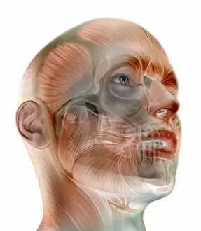
Background imageOral Collection: Human facial muscles, artwork

Human facial muscles, artwork
Human facial muscles. Artwork of the facial and other muscles of a human head and neck, seen from an oblique frontal angle

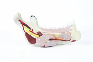
Background imageOral Collection: Human lower jaw, historical model

Human lower jaw, historical model
Human lower jaw. 1950s model showing the structure of the lower jaw (mandible) of a human

Background imageOral Collection: Human tooth, historical anatomical model

Human tooth, historical anatomical model
Human tooth. 1950s model showing the external structure of a human molar tooth

Background imageOral Collection: Earthworm head, SEM

Earthworm head, SEM
Earthworm head. Coloured scanning electron micrograph (SEM) of the head of a small annelid (segmented) earthworm, showing its mouth, and small clumps of setae (bristles)

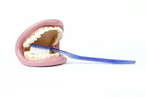
Background imageOral Collection: Human mouth, anatomical model

Human mouth, anatomical model
Human mouth. Anatomical model (from 1995) of a human mouth, showing the lips (pink), gums (gingiva, beige), and teeth (white) - being brushed with a toothbrush

Background imageOral Collection: Mouth cancer cells, SEM

Mouth cancer cells, SEM
Mouth cancer cells. Coloured scanning electron micrograph (SEM) of squamous cell carcinoma (cancer) cells from a human mouth

Background imageOral Collection: Velvet skull, anatomical model

Velvet skull, anatomical model
Velvet skull. Velvet-covered anatomical model of a human skull

Background imageOral Collection: Mouth cancer cell dividing, SEM

Mouth cancer cell dividing, SEM
Mouth cancer dividing. Coloured scanning electron micrograph (SEM) of a squamous cell carcinoma (cancer) cell from a human mouth undergoing mitosis (nuclear division)

Background imageOral Collection: Mouth cancer cell, SEM

Mouth cancer cell, SEM
Mouth cancer cell. Coloured scanning electron micrograph (SEM) showing the surface of a squamous cell carcinoma (cancer) cell from a human mouth

Background imageOral Collection: Human jaw, 19th Century model

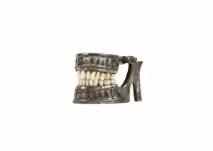
Human jaw, 19th Century model. 1870 dental teaching aid showing the structure of the human jaws

Background imageOral Collection: Mouth, artwork

Mouth, artwork
Mouth. Computer artwork showing the anatomy of the human mouth. Seen here are the teeth (white), tongue (lower centre), uvula (centre), and tonsils (red, centre left and right)

Background imageOral Collection: Dental hygiene set, circa 1870 C017 / 8407

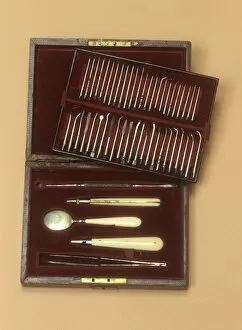
Dental hygiene set, circa 1870 C017 / 8407
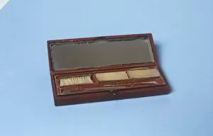
Comprehensive dental hygiene set in a morocco case. The set includes an inspection mirror, steel tools, and ivory handles

Background imageOral Collection: Two folding toothpicks, circa 1890 C017 / 8405

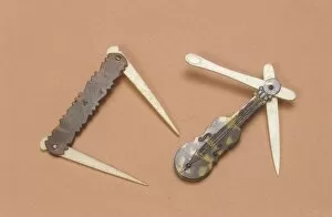
Two folding toothpicks, circa 1890 C017 / 8405
Two folding toothpicks, dating from around 1890. The left features two pointed toothpicks. The right is a novelty violin shape in faux tortoiseshell and has one folding toothpick and one ear scoop

Background imageOral Collection: Four tongue scrapers, 19th century C017 / 8404

Four tongue scrapers, 19th century C017 / 8404
Four assorted tongue scrapers dating from between 1800 and 1850, England. These are made out of silver, ivory and tortoiseshell

Background imageOral Collection: Four tongue scrapers, circa 1850 C017 / 8400

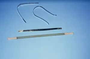
Four tongue scrapers, circa 1850 C017 / 8400
Four tongue scrapers, three in silver and one in ivory. These date from around 1850, England. Tongue scrapers such as these were hand crafted personal hygiene instruments

Background imageOral Collection: Spiral mouth gag, circa 1860 C017 / 8396

Spiral mouth gag, circa 1860 C017 / 8396
Spiral mouth gag, made from boxwood, dating from around 1860, England. This would have been used to pry open the mouths of patients with lock-jaw

Background imageOral Collection: Dental saw, circa 1870 C017 / 8399

Dental saw, circa 1870 C017 / 8399
Dental saw with ebony handle dating from around 1870, England. Dental saws were used in early dentistry to create separations between the teeth

Background imageOral Collection: Dental compendium, circa 1860 C017 / 8398

Dental compendium, circa 1860 C017 / 8398
Comprehensive dental compendium, complete with forceps, tooth keys, elevators, and dental picks. The nineteenth century was a period of rapid progression in the field of dentistry

Background imageOral Collection: Six dental picks, circa 1860 C017 / 8386

Six dental picks, circa 1860 C017 / 8386
Six dental picks, circa 1860

Background imageOral Collection: Mouth gag, circa 1870 C017 / 8385

Mouth gag, circa 1870 C017 / 8385
Steel mouth gag with a cross hatched ebony handle. This would have been used to pry open the mouths of patients with lock-jaw or to hold the jaw in position for tooth extraction

Background imageOral Collection: Tongue scraper, circa 1850 C017 / 8377

Tongue scraper, circa 1850 C017 / 8377
Ivory and tortoiseshell tongue scraper. Tongue scrapers such as this one were handcrafted personal hygiene instruments, specially designed for the removal of debris which build up on the tongue

Background imageOral Collection: Six dental picks, circa 1860 C017 / 8379

Six dental picks, circa 1860 C017 / 8379
Six steel dental picks with ivory handles. The nineteenth century was a period of rapid progression in the field of dentistry

Background imageOral Collection: Dental hygiene set, circa 1821 C017 / 8373

Dental hygiene set, circa 1821 C017 / 8373
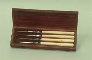
Four steel dental hygiene tools with ivory handles dating from around 1820, England. These are shown in their original morocco case

Background imageOral Collection: Tongue scrapers, circa 1850 C017 / 8376

Tongue scrapers, circa 1850 C017 / 8376
Two ivory and tortoiseshell tongue scrapers. These were handcrafted personal hygiene instruments, specially designed for the removal of debris which build up on the tongue

Background imageOral Collection: Dental hygiene set, circa 1820 C017 / 8367

Dental hygiene set, circa 1820 C017 / 8367
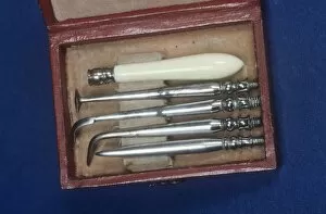
Boxed dental hygiene set with four steel tools and a detachable ivory handle. This set is English and dates from around 1820

Background imageOral Collection: Six tongue scrapers, 19th century C017 / 8369

Six tongue scrapers, 19th century C017 / 8369
Six tongue scrapers in ivory, tortoiseshell and silver. These were handcrafted personal hygiene instruments, specially designed for the removal of debris which build up on the tongue

Background imageOral Collection: Two dental mirrors, circa 1870 C017 / 8366

Two dental mirrors, circa 1870 C017 / 8366
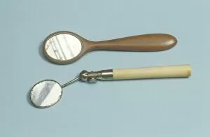
Two concave mirrors for dental hygiene, one is made from ivory and the other from wood. The nineteenth century was a period of rapid progression in the field of dentistry and dental hygiene

Background imageOral Collection: Cased toilet set, circa 1800 C017 / 8361

Cased toilet set, circa 1800 C017 / 8361
Cased toilet set dating from around 1800, England. The set contains a toothbrush, tooth powder box, silver tongue scraper and toothpick

Background imageOral Collection: Ear scoop and toothpicks, circa 1890 C017 / 8360

Ear scoop and toothpicks, circa 1890 C017 / 8360
Two toothpicks, the lower one is in silver and the upper with folding mechanism and ear scoop. These date from around 1890, England

Background imageOral Collection: Dental X-rays

Dental X-rays

Background imageOral Collection: Dental inspection C015 / 3452

Dental inspection C015 / 3452
Dental inspection. Close-up of a patients teeth being inspected with a mirror

Background imageOral Collection: Fluoride in toothpaste, conceptual image C017 / 8045

Fluoride in toothpaste, conceptual image C017 / 8045
Fluoride in toothpaste, conceptual computer artwork

Background imageOral Collection: Osteonecrosis of the jaw, artwork C017 / 8039

Osteonecrosis of the jaw, artwork C017 / 8039
Osteonecrosis of the jaw. Computer artwork showing osteonecrosis (death of bone tissue) in a persons right mandible (lower jaw bone)

Background imageOral Collection: Dentist

Dentist
MODEL RELEASED. Dentist. Close-up of a dental mirror being held by a dentist

Background imageOral Collection: Dental model

Dental model. Close-up of a clay cast of a human mouth with an artificial resin tooth (centre right)

Background imageOral Collection: Dental cast

Dental cast. Close-up of a cast of a human tooth

Background imageOral Collection: Deaf-mute education, 1917 C016 / 4516

Deaf-mute education, 1917 C016 / 4516
Deaf-mute education. Teacher teaching a deaf-mute girl to talk. The girl has her hand placed on the teachers throat to feel the sounds made while talking

Background imageOral Collection: Bexarotene chemotherapy drug molecule C013 / 7785

Bexarotene chemotherapy drug molecule C013 / 7785
Bexarotene chemotherapy drug molecule. Computer model showing the structure of a molecule of the antineoplastic drug bexarotene (C24H28O2)


For sale as Licensed Images

Choose your image, Select your licence and Download the media

"Exploring the Intricacies of Oral Health: From Ancient Art to Modern Science" Step into the world health as we unravel its fascinating secrets through a diverse range of captivating images. Leonardo da Vinci's meticulous "Skull anatomy" drawing takes us back in time, showcasing his unparalleled understanding of dental structures. A panoramic dental X-ray reveals a comprehensive view, allowing dentists to diagnose and treat conditions with precision. Meanwhile, in Senegal, a Griot skillfully plays the Kora, reminding us of the cultural significance attached to oral traditions and storytelling. False teeth serve as a testament to mankind's quest for restoring smiles and functionality. A polarised LM image showcases decay within a molar tooth, highlighting the importance of regular check-ups and proper oral hygiene practices. The intricate illustration depicting our respiratory system reminds us that our oral cavity is not isolated but intricately connected to our overall well-being. Understanding this connection helps prevent various diseases affecting both our mouth and lungs. Delving deeper into dental knowledge, an illustration unveils the complex anatomy behind our teeth – each playing an essential role in chewing and speech production. Used dental floss under SEM magnification emphasizes the significance of interdental cleaning for maintaining optimal oral health. An X-ray revealing a fractured jawbone serves as a reminder that accidents can impact not only aesthetics but also functional aspects of one's smile. However, advancements like Januvia diabetes drug molecule offer hope by addressing underlying conditions affecting oral health. Zooming closer into modern dentistry marvels, an LM cross-section showcases how dental implants seamlessly integrate with surrounding tissues – revolutionizing tooth replacement options for countless individuals worldwide. Lastly, we witness everyday care routines as a man diligently brushes his teeth – reinforcing that simple habits lay the foundation for lifelong oral wellness. In this journey through artistry and science intertwined with human experiences across cultures and time periods, it becomes evident that caring for your mouth goes beyond just a smile – it is an essential part of overall health and well-being.