
Organs Collection (#8)
"Theosophy Chakras 1696: Exploring the Mystical Connection between Organs and Energy Centers" Delve into the intricate world as we unravel their hidden secrets


For sale as Licensed Images
Choose your image, Select your licence and Download the media
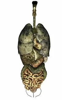
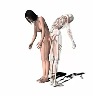
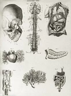
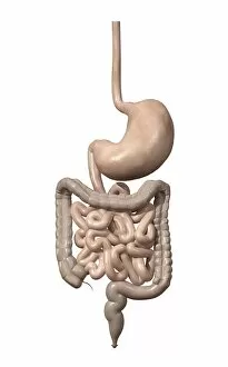
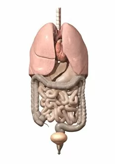
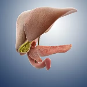
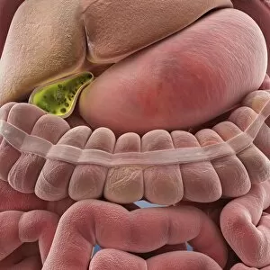
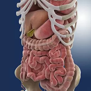
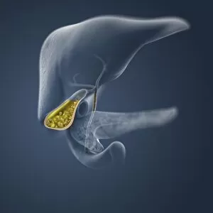
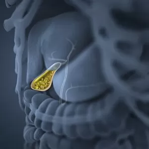
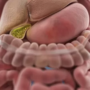
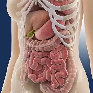
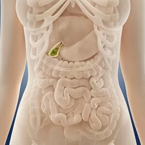
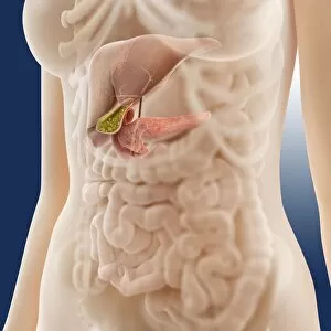
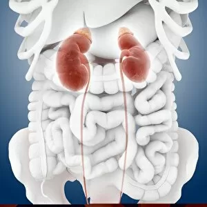
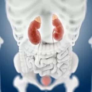
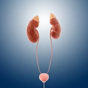
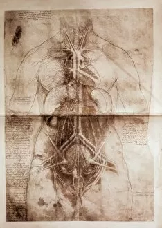
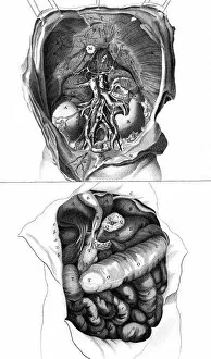
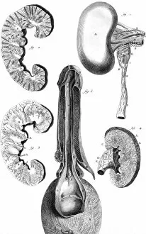
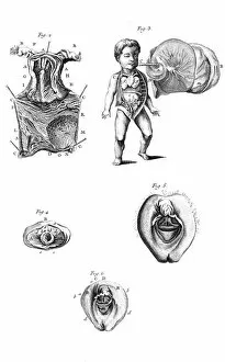
"Theosophy Chakras 1696: Exploring the Mystical Connection between Organs and Energy Centers" Delve into the intricate world as we unravel their hidden secrets. From the Front View of Lungs to a Diagram showcasing their complex structure, witness the marvels that keep us breathing. Discover the Master organ at Saint-Eustache church in Paris, France, Europe - an architectural wonder intertwined with spirituality. Marvel at historical artwork depicting the Cardiovascular system, offering glimpses into our ancient understanding of these vital organs. Uncover surprising connections between Astrology and medicine through captivating artwork. Explore Neck anatomy through mesmerizing 19th Century illustrations that showcase both beauty and complexity. Step back in time to witness an Automaton at the Schoolboys Own Exhibition in 1929, a testament to human ingenuity mimicking our own organic machinery. Immerse yourself in artistic depictions of Pig anatomy, where science meets creativity. Witness various parts come together harmoniously within Anatomy's intricate web. Embark on a journey through Digestive Organs - from mouth-watering beginnings to nourishing endings. Understand how each piece plays its unique role in this complex symphony of digestion. Lastly, explore Phrenological Head captured in black and white - a fascinating study intertwining psychology and physiology for centuries. Join us as we unlock the mysteries behind these remarkable organs that sustain life itself.