
Orthopedic Collection

"Exploring the World of Orthopedic: From Damaged Knee Ligaments to Artwork" Orthopedics

Background imageOrthopedic Collection: Headache, X-ray artwork

Headache, X-ray artwork
Headache. Computer X-ray artwork of a lateral view of a human skull. The red area represents pain caused by a headache (cephalalgia)

Background imageOrthopedic Collection: Damaged knee ligament, artwork

Damaged knee ligament, artwork
Damaged knee ligament. Computer artwork of the muscle structure of the leg and anatomy of the human knee joint, showing a tear (dark line, centre right) in one of the cruciate ligaments (beige)

Background imageOrthopedic Collection: Fractured jawbone, X-ray

Fractured jawbone, X-ray
Fractured jawbone. Coloured X-ray of a human jaw showing a fractured lower jawbone (mandible). The fracture has occurred to the left side of the lower jawbone (bottom right, orange)

Background imageOrthopedic Collection: Treatment of a dislocation, from a commentary of Apollonius of Citium (1st century BC

Treatment of a dislocation, from a commentary of Apollonius of Citium (1st century BC
CHT236783 Treatment of a dislocation, from a commentary of Apollonius of Citium (1st century BC) on the Hippocratic treatise On Joints (vellum) by Greek School

Background imageOrthopedic Collection: Broken arm bone, digital X-ray

Broken arm bone, digital X-ray
Broken arm bone. Digital frontal X-ray of the fractured right humerus (upper arm bone) of a patient in their early twenties

Background imageOrthopedic Collection: Normal hand, digital X-ray

Normal hand, digital X-ray
Normal hand. Digital X-ray of the normal right hand of a patient in their late twenties. Various sets of data relating to the image can be seen around the edges of the X-ray

Background imageOrthopedic Collection: Anatomy of hip fracture

Anatomy of hip fracture

Background imageOrthopedic Collection: Taped sprained ankle C017 / 3695

Taped sprained ankle C017 / 3695
Taped sprained ankle. Close-up of elastic therapeutic tape (kinesio tape) being used to promote lymphatic drainage, and therefore reduce swelling, in a womans sprained ankle

Background imageOrthopedic Collection: Amputated toes, X-ray C017 / 7166

Amputated toes, X-ray C017 / 7166
Amputated toe. X-ray of the left foot of a 75 year old male patient where the tip of the big toe and two thirds of the next toe have been amputated

Background imageOrthopedic Collection: Amputated fingers, X-ray C017 / 7150

Amputated fingers, X-ray C017 / 7150
Amputated fingers. X-ray of the right hand of a 57 year old male patient where the tips of the fourth and fifth fingers have been amputated

Background imageOrthopedic Collection: Radiologist examining knee MRI scans C014 / 1300

Radiologist examining knee MRI scans C014 / 1300
MODEL RELEASED. Radiologist examining knee MRI (magnetic resonance imaging) scans

Background imageOrthopedic Collection: Tuberculosis of the spine, MRI scan

Tuberculosis of the spine, MRI scan
Tuberculosis of the spine. Magnetic resonance imaging (MRI) scan of a sagittal section through the spine of a 74-year-old patient with tuberculosis (TB) of the spine, or Potts disease

Background imageOrthopedic Collection: Spondylolisthesis, 3D CT scan

Spondylolisthesis, 3D CT scan
Spondylolisthesis. Coloured 3D computed tomography (CT) scan of a section through the lumbar (lower back) spine of a 39-year-old patient with spondylolisthesis

Background imageOrthopedic Collection: Paprosky femur defect classification C016 / 6621

Paprosky femur defect classification C016 / 6621
Paprosky femur defect classification. Diagram showing the classification system for femur cortex defects used to assess revision (replacement or repair) of a hip implant

Background imageOrthopedic Collection: Coccyx pain, artwork

Coccyx pain, artwork
Coccyx (tailbone) pain, computer artwork

Background imageOrthopedic Collection: Neck and shoulder pain, X-ray artwork

Neck and shoulder pain, X-ray artwork
Neck and shoulder pain. Computer X-ray artwork of the back view of a human skeletons shoulders, neck and skull. The red area represents neck pain (cervicalgia) and shoulder pain

Background imageOrthopedic Collection: Detail of human knee showing insertion of arthroscopic instruments

Detail of human knee showing insertion of arthroscopic instruments

Background imageOrthopedic Collection: Head of femur fracture

Head of femur fracture

Background imageOrthopedic Collection: Detail of human knee showing arthroscopic surgical proceedures

Detail of human knee showing arthroscopic surgical proceedures

Background imageOrthopedic Collection: USS COMFORT, c1919. Orthopedic operating room aboard the hospital ship USS Comfort

USS COMFORT, c1919. Orthopedic operating room aboard the hospital ship USS Comfort. Photograph, c1919

Background imageOrthopedic Collection: Scoliosis of the spine, X-ray C017 / 7167

Scoliosis of the spine, X-ray C017 / 7167
Scoliosis. X-ray of the spine of a 71 year old female patient with severe scoliosis (sideways curvature). This condition usually starts in infancy or adolescence, and is most often seen in girls

Background imageOrthopedic Collection: Melorheostosis of the knee, X-ray C017 / 7146

Melorheostosis of the knee, X-ray C017 / 7146
Melorheostosis of the knee. X-ray of left knee of a 52 year old male patient showing damage (white area at centre) caused by melorheostosis

Background imageOrthopedic Collection: Melorheostosis of the knee, X-ray C017 / 7145

Melorheostosis of the knee, X-ray C017 / 7145
Melorheostosis of the knee. X-ray of left knee of a 52 year old male patient showing damage (white area to bottom left of knee cap) caused by melorheostosis

Background imageOrthopedic Collection: Melorheostosis of the knee, X-ray C017 / 7144

Melorheostosis of the knee, X-ray C017 / 7144
Melorheostosis of the knee. X-ray of left knee of a 52 year old male patient showing damage (white area to bottom left of knee cap) caused by melorheostosis

Background imageOrthopedic Collection: Hip pain, artwork C013 / 5917

Hip pain, artwork C013 / 5917
Hip pain, computer artwork

Background imageOrthopedic Collection: Sciatica, MRI scan

Sciatica, MRI scan
Sciatica. Coloured magnetic resonance imaging (MRI) scan of a sagittal section through the lumbar (lower back) spine of a 35-year-old patient

Background imageOrthopedic Collection: Spondylodiscitis, MRI scan

Spondylodiscitis, MRI scan
Spondylodiscitis. Coloured magnetic resonance imaging (MRI) scan of a sagittal section through the spine of a 69-year-old patient with spondylodiscitis at the level of the T7-T8 vertebra

Background imageOrthopedic Collection: Abnormal bone growth, MRI scan

Abnormal bone growth, MRI scan
Abnormal bone growth. Magnetic resonance imaging (MRI) scan of a section through the cervical spine (neck) of a 28-year-old patient, showing abnormal growth (dysplasia) of the C6 vertebra

Background imageOrthopedic Collection: Spinal cancer, MRI scan

Spinal cancer, MRI scan
Spinal cancer. Coloured magnetic resonance imaging (MRI) scan of a sagittal section through the cervical (neck) region of the spine of a 22-year-old patient

Background imageOrthopedic Collection: Forestiers disease, MRI scan

Forestiers disease, MRI scan
Forestiers disease. Magnetic resonance imaging (MRI) scan of a section through the cervical spine (neck) of a 42-year-old Asian patient suffering from a stiff neck and neck pain

Background imageOrthopedic Collection: Spina bifida, MRI scan

Spina bifida, MRI scan
Fibrolipoma of the spine. Magnetic resonance imaging (MRI) scan of a sagittal section through the spine of a 24-month-old patient with spina bifida

Background imageOrthopedic Collection: Secondary spinal cancer, MRI scan

Secondary spinal cancer, MRI scan
Secondary spinal cancer. Magnetic resonance imaging (MRI) scan of a section through the spine of a 48-year-old patient, showing malignant (cancerous) tumours

Background imageOrthopedic Collection: Fibrolipoma of the spine, MRI scan

Fibrolipoma of the spine, MRI scan
Fibrolipoma of the spine. Coloured magnetic resonance imaging (MRI) scan of a sagittal section through the thoraco-lumbar spine of a 19-month-old patient

Background imageOrthopedic Collection: Total knee replacement, X-ray

Total knee replacement, X-ray
Total knee replacement. X-ray of the legs of a 49-year-old patient who has had a total knee replacement (white) of the left knee (right)

Background imageOrthopedic Collection: Prosthetic hip joint and Gruen zones C016 / 6778

Prosthetic hip joint and Gruen zones C016 / 6778
Prosthetic hip joint and Gruen zones. Cutaway diagram of a femur (thigh bone) showing a femoral component of a hip prosthesis and labels indicating Gruen zones

Background imageOrthopedic Collection: Prosthetic hip joints and Gruen zones C016 / 6781

Prosthetic hip joints and Gruen zones C016 / 6781
Prosthetic hip joints and Gruen zones. Diagram of the femoral components of two types of hip prosthesis with labels indicating standard and modified Gruen zones

Background imageOrthopedic Collection: Prosthetic hip joint, artwork C016 / 6782

Prosthetic hip joint, artwork C016 / 6782
Prosthetic hip joint. Cutaway artwork of a femur (thigh bone) showing a femoral component of a hip prosthesis. This component is implanted in the femur after the head of the femur has been surgically

Background imageOrthopedic Collection: Hip socket bone grafting, diagram C016 / 6786

Hip socket bone grafting, diagram C016 / 6786
Hip socket bone grafting. Diagram showing completed hip socket bone grafting as part of a total hip replacement operation

Background imageOrthopedic Collection: Hip socket bone grafting, diagram C016 / 6784

Hip socket bone grafting, diagram C016 / 6784
Hip socket bone grafting. Diagram showing the second stage in hip socket bone grafting as part of a total hip replacement operation

Background imageOrthopedic Collection: Hip socket bone grafting, diagram C016 / 6785

Hip socket bone grafting, diagram C016 / 6785
Hip socket bone grafting. Diagram showing the third stage in hip socket bone grafting as part of a total hip replacement operation

Background imageOrthopedic Collection: Hip socket bone grafting, diagram C016 / 6783

Hip socket bone grafting, diagram C016 / 6783
Hip socket bone grafting. Diagram showing the first stage in hip socket bone grafting as part of a total hip replacement operation

Background imageOrthopedic Collection: Prosthetic hip joint, diagram C016 / 6775

Prosthetic hip joint, diagram C016 / 6775
Prosthetic hip joint. Cutaway diagram of a femur (thigh bone) showing a femoral component of a hip prosthesis. This component is implanted in the femur after the head of the femur has been surgically

Background imageOrthopedic Collection: Prosthetic hip joint and Gruen zones C016 / 6777

Prosthetic hip joint and Gruen zones C016 / 6777
Prosthetic hip joint and Gruen zones. Cutaway diagram of a femur (thigh bone) showing a femoral component of a hip prosthesis and labels indicating Gruen zones

Background imageOrthopedic Collection: Prosthetic hip joint and Gruen zones C016 / 6780

Prosthetic hip joint and Gruen zones C016 / 6780
Prosthetic hip joint and Gruen zones. Diagram of the femoral component of a hip prosthesis with labels indicating the hip connection (upper left) and the seven Gruen zones

Background imageOrthopedic Collection: Prosthetic hip joint and Gruen zones C016 / 6776

Prosthetic hip joint and Gruen zones C016 / 6776
Prosthetic hip joint and Gruen zones. Cutaway diagram of a femur (thigh bone) showing a femoral component of a hip prosthesis and labels indicating Gruen zones

Background imageOrthopedic Collection: Prosthetic hip joint and Gruen zones C016 / 6779

Prosthetic hip joint and Gruen zones C016 / 6779
Prosthetic hip joint and Gruen zones. Cutaway diagram of a femur (thigh bone) showing a femoral component of a hip prosthesis and labels indicating Gruen zones

Background imageOrthopedic Collection: Total hip replacement, X-rays

Total hip replacement, X-rays
Total hip replacement. Coloured X-rays taken during (left) and after (right) surgery to replace the right hip joint of a 65-year-old patient using a prosthetic implant

Background imageOrthopedic Collection: Failed hip replacement, CT scan

Failed hip replacement, CT scan
Failed hip replacement. Coloured computed tomography (CT) scan of a total hip replacement (solid white) in a 70-year-old patient


For sale as Licensed Images

Choose your image, Select your licence and Download the media

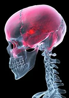
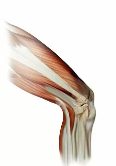
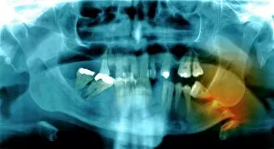
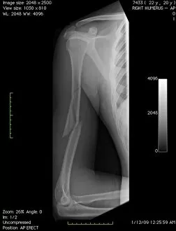
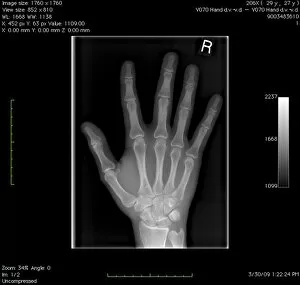
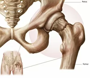
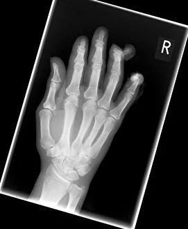
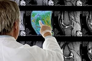
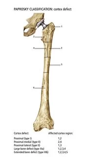
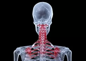
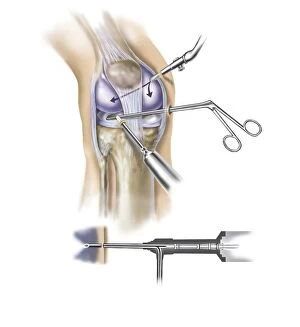
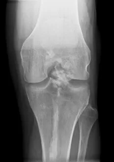
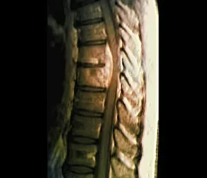
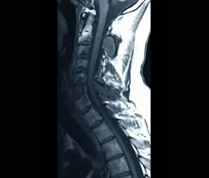
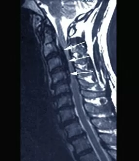
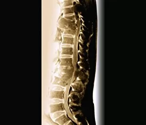
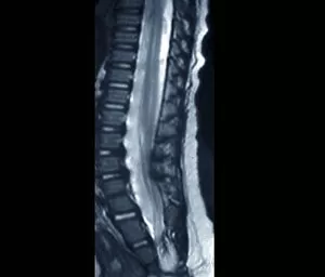
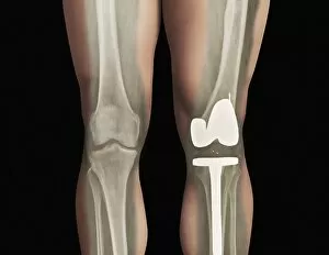
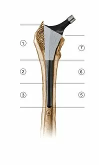
"Exploring the World of Orthopedic: From Damaged Knee Ligaments to Artwork" Orthopedics, a fascinating field that delves into the intricate workings of our musculoskeletal system. It encompasses a wide range of conditions and treatments, each with its own unique story to tell. Let's start with a damaged knee ligament, where pain meets artistry. The delicate balance between injury and healing is captured in an artwork that portrays both vulnerability and resilience. Moving on to headaches, we delve deeper through X-ray artwork. A glimpse into the inner workings of our skulls reveals the hidden battles within, as throbbing pain takes center stage amidst intricate bone structures. Fractured jawbones come next, their stories etched onto X-ray films. These images speak volumes about resilience and recovery as shattered bones mend together again, forming a stronger foundation than before. Traveling back in time to ancient wisdom, we find ourselves immersed in Apollonius of Citium's commentary on treating dislocations. His words echo through centuries as we witness the evolution care from ancient practices to modern techniques. A broken arm bone comes alive through digital X-rays - a testament to technological advancements aiding diagnosis and treatment plans. These images remind us that even in moments of fragility, there is hope for restoration and renewal. Shifting focus towards normalcy, let's explore the beauty hidden within our hands using digital X-rays. Delicate bones intertwine like pieces of artistry themselves - showcasing nature's design at its finest. Jean-Jacques Manget's Bibliotheca guides us further into this captivating realm. Within its pages lie treasures waiting to be discovered - knowledge passed down by generations who dedicated their lives to understanding orthopedics' intricacies. Anatomy takes center stage once more as we unravel the secrets behind hip fractures. Through detailed illustrations and explanations, we gain insight into how these injuries occur and the path to recovery that lies ahead.