
Orthopedics Collection
Orthopedics, the branch of medicine that focuses on treating injuries and disorders of the musculoskeletal system


For sale as Licensed Images
Choose your image, Select your licence and Download the media
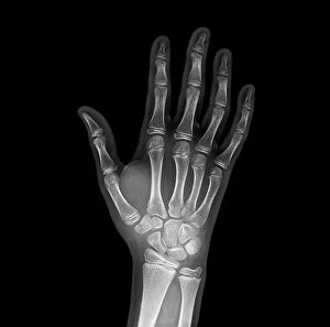
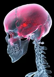
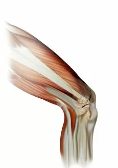
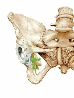
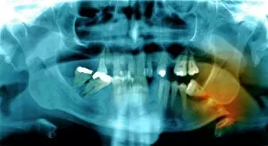
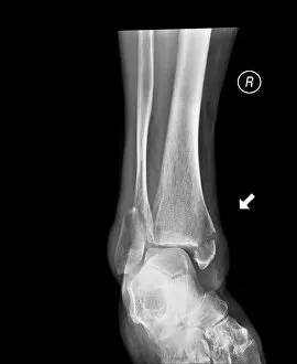
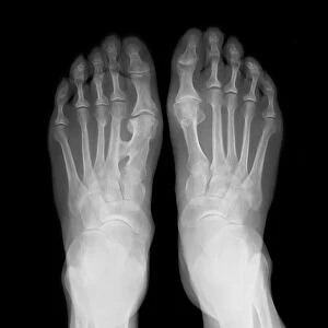
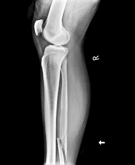
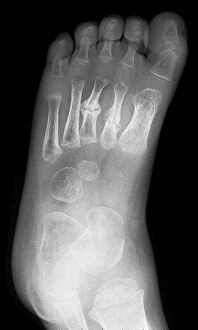
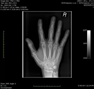
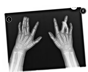
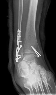
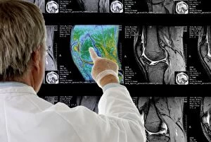
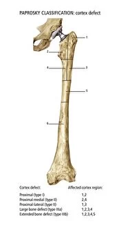
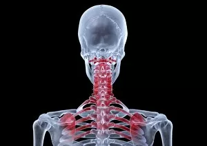
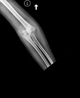
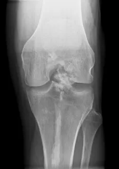
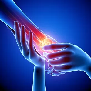
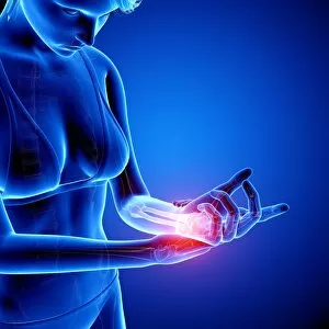
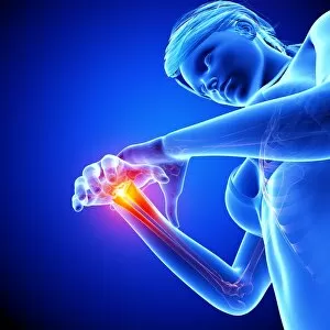
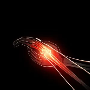
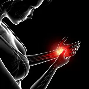
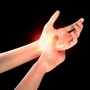
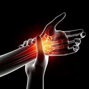
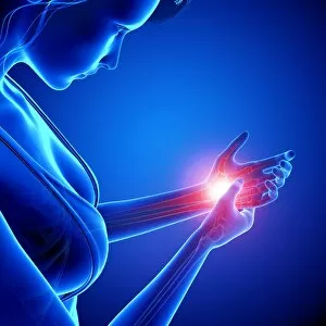
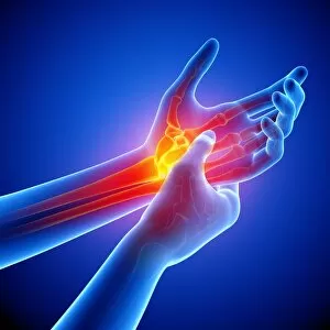
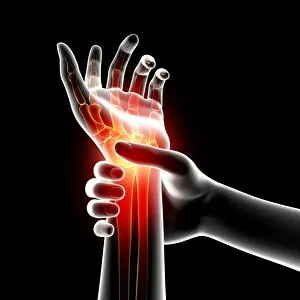
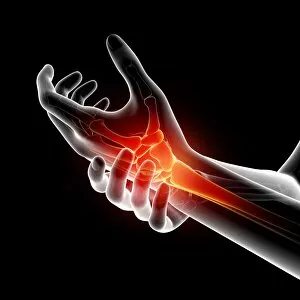
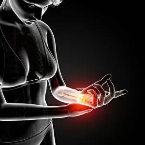
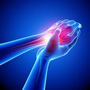
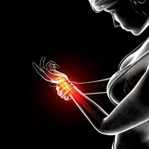
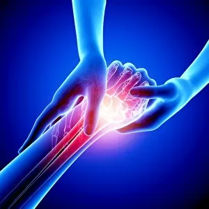
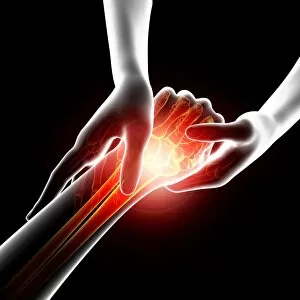
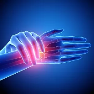
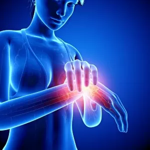
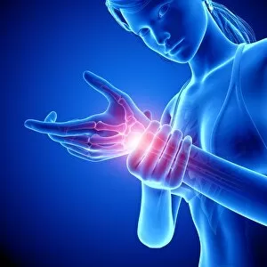
Orthopedics, the branch of medicine that focuses on treating injuries and disorders of the musculoskeletal system, plays a crucial role in restoring mobility and improving quality of life. From broken wrist bones to damaged knee ligaments, orthopedic specialists are skilled in diagnosing and treating a wide range of conditions. In the realm of orthopedics, technology has become an invaluable tool. X-rays like C017 / 7187 reveal intricate details about fractures such as a broken wrist bone or a fractured jawbone. These images guide physicians in devising effective treatment plans tailored to each patient's needs. Artwork also serves as an unconventional medium for showcasing orthopedic challenges. A piece depicting a damaged knee ligament reminds us of the importance of proper care and rehabilitation after injury. Similarly, an artwork portraying a hip replacement highlights how advancements in surgical techniques have revolutionized joint replacements. The history is rich with notable figures who have contributed significantly to this field. Welsh surgeon Hugh Owen Thomas (C016 / 6299) stands out as one such pioneer whose innovative methods shaped modern orthopedic practices. X-ray images continue to be instrumental in diagnosing various conditions affecting different parts of our body. Whether it's identifying fractures like those seen in X-rays C017 / 7185 or C017 / 7977 or detecting abnormalities like fused metatarsals (X-ray C017 / 7168), these diagnostic tools aid doctors in providing accurate diagnoses and appropriate treatments. Not all X-rays depict injuries; some showcase normal anatomy too. A digital X-ray displaying a healthy hand reminds us that not every visit to an orthopedist involves trauma but can involve routine check-ups or preventive measures. Lastly, we encounter cases where even our vertebrae may suffer fractures (X-ray C017 / 7586). Orthopedists play a vital role here by offering solutions that alleviate pain while promoting healing and recovery.