
Ossification Collection
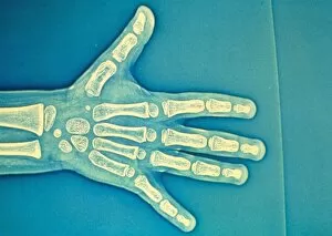
"Unveiling the Journey of Ossification: From Fragility to Strength" A glimpse into growth: Exploring ossification through a child hand X-ray


For sale as Licensed Images
Choose your image, Select your licence and Download the media
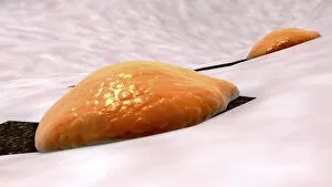
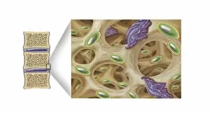
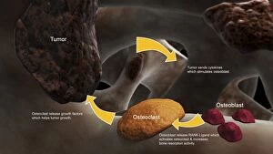
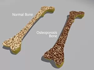
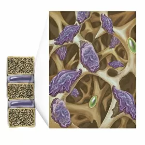
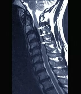
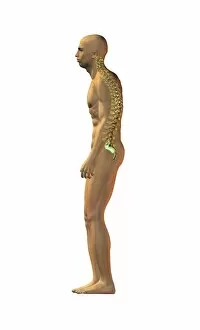
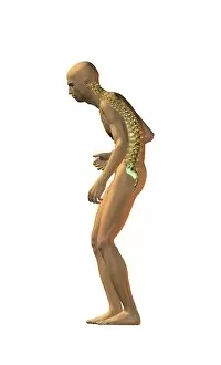
"Unveiling the Journey of Ossification: From Fragility to Strength" A glimpse into growth: Exploring ossification through a child hand X-ray. Unraveling bone remodeling: Witness osteoclasts breaking down bone tissue. Building blocks of strength: Discover how osteoblasts construct healthy bones. The invasive spread: Conceptual image depicting the process of bone metastasis. A tale of two bones: Comparing a healthy bone with one affected by osteoporosis. Weakening foundations: Osteoclasts eroding bones in the battle against osteoporosis. Peering into Forestier's disease: Insights from an MRI scan revealing spinal abnormalities. Embracing normality: Appreciating artwork showcasing a spine's ideal posture. Onset of spinal disorders: Artwork capturing the moment when ailments take hold. Unmasking pain and tension: Visualizing discomfort through artistic representation. Navigating spinal disorder complexities through artistry and creativity. Embark on a visual journey that explores ossification, where fragile bones transform into pillars of strength and resilience. Delve deep into this captivating process as we begin with an intimate look at a child's hand X-ray, unveiling the intricate world within. Witness the relentless work of osteoclasts as they break down old or damaged bone tissue, paving the way for renewal and rejuvenation in our bodies' skeletal framework. But it doesn't end there - meet our unsung heroes, osteoblasts. These remarkable cells diligently build healthy new bone structures, ensuring our bodies remain strong and functional throughout life's challenges. Yet sometimes, this delicate balance is disrupted by unforeseen circumstances like bone metastasis - an invasive journey that threatens to compromise our very foundation. Take a closer look at two contrasting realities.