
Osteological Collection
"Exploring the Intricacies of Osteology: From Fractured Ankles to Rare Bone Disorders" Step into the world of osteology


For sale as Licensed Images
Choose your image, Select your licence and Download the media
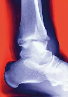
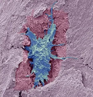
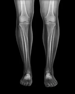
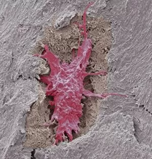
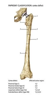
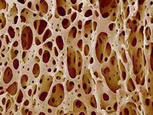
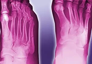
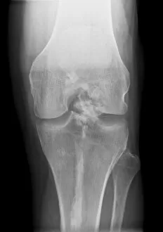
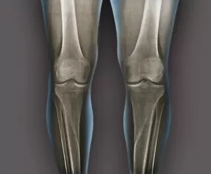
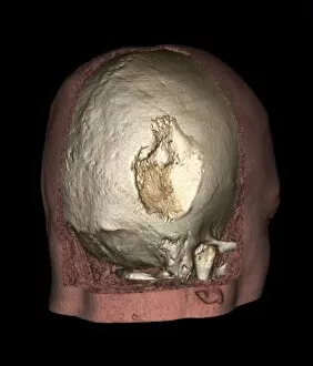
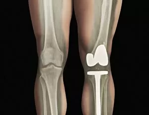
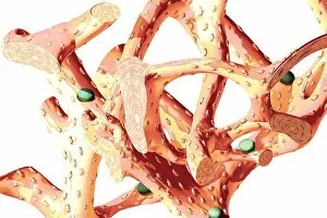
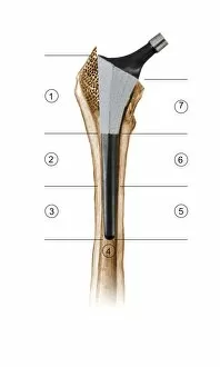
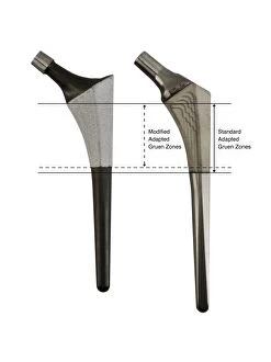
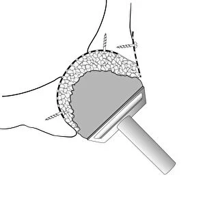
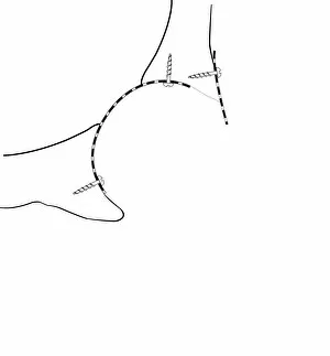
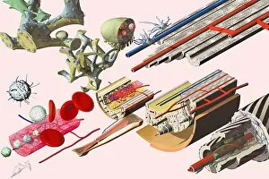
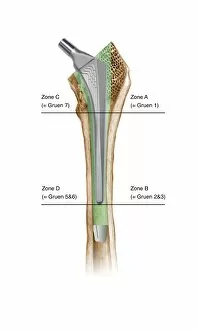
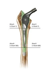
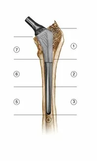
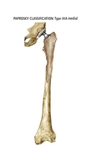
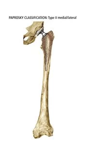
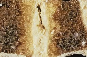
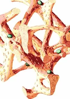
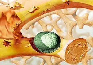
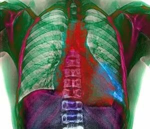
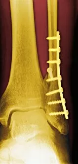
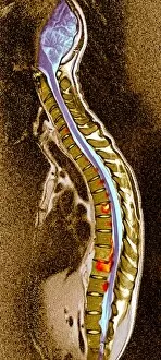
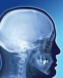
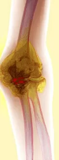
"Exploring the Intricacies of Osteology: From Fractured Ankles to Rare Bone Disorders" Step into the world of osteology, where bones reveal their secrets through various hints and images. In a dimly lit room at the College of Surgeons in the 1880s, anatomists meticulously studied fractured ankles captured on X-rays. These haunting images offered glimpses into the intricate structure of our skeletal system. Zooming in further, we encounter osteocyte bone cells under an electron microscope (SEM C016 / 9025). These tiny warriors play a vital role in maintaining bone health and repair. Fast forward to July 5th, 1892, when an awe-inspiring Osteological Gallery opened its doors. Visitors marveled at displays showcasing normal legs captured by X-rays - a testament to the marvels hidden beneath our skin. Delving deeper into cellular wonders, another SEM image (C016 / 9026) reveals more about osteocytes' microscopic world. Their interconnected network ensures strength and resilience within our bones. But not all is smooth sailing; challenges arise like Paprosky femur defect classification (C016 / 6621), which helps surgeons navigate complex bone issues with precision and expertise. The enigmatic Melorheostosis makes its appearance through X-ray images (C017 / 7146-7144). This rare condition causes abnormal growth patterns within bones, posing unique diagnostic challenges for medical professionals. Returning to normalcy, we witness yet again how X-rays shed light on healthy knees - providing baseline comparisons for diagnosing ailments that affect this crucial joint's functionality. Lastly, Gorham's disease takes center stage as a three-dimensional CT scan unravels its mysteries. This rare disorder leads to progressive loss of bone tissue and serves as a reminder of nature's unpredictability. Osteological studies offer us profound insights into our skeletal framework – from fractures to rare conditions.