Oviduct Collection

The oviduct, also known as the fallopian tube, is a crucial part of the female reproductive system

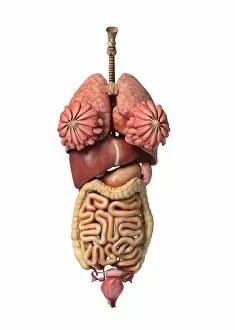
Background imageOviduct Collection: 3D rendering of healthy female internal organs

3D rendering of healthy female internal organs

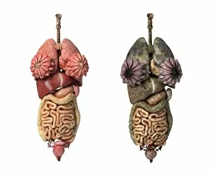
Background imageOviduct Collection: Comparison of healthy and unhealthy female organs

Comparison of healthy and unhealthy female organs
3D rendering comparing healthy female organs and unhealthy female organs

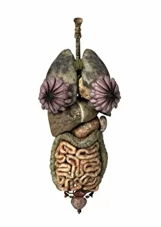
Background imageOviduct Collection: 3D rendering of unhealthy female internal organs

3D rendering of unhealthy female internal organs

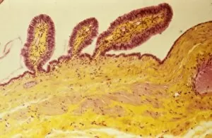
Background imageOviduct Collection: Fallopian tube, light micrograph F006 / 9799

Fallopian tube, light micrograph F006 / 9799
Fallopian tube. Light micrograph of a cross-section through a fallopian tube. The lumen (white) is seen, along with mucosal folds lined with simple columnar ciliated epithelium (purple)

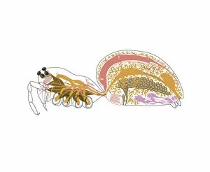
Background imageOviduct Collection: Spider anatomy, artwork

Spider anatomy, artwork
Spider anatomy. Computer artwork showing the main organs of a typical female spider

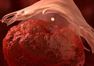
Background imageOviduct Collection: Conceptual image of sperm inside fallopian tube

Conceptual image of sperm inside fallopian tube

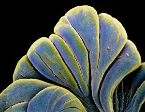
Background imageOviduct Collection: Fimbriae of a fallopian tube, SEM C016 / 9047

Fimbriae of a fallopian tube, SEM C016 / 9047
Fimbriae of a fallopian tube, coloured scanning electron micrograph (SEM). Fimbriae are the folds at the opening (ostium) of a fallopian tube, next to an ovary

Background imageOviduct Collection: Ovulation, artwork C016 / 7537

Ovulation, artwork C016 / 7537
Ovulation, computer artwork

Background imageOviduct Collection: Fimbriae of a fallopian tube, SEM C016 / 9048

Fimbriae of a fallopian tube, SEM C016 / 9048
Fimbriae of a fallopian tube, coloured scanning electron micrograph (SEM). Fimbriae are the folds at the opening (ostium) of a fallopian tube, next to an ovary

Background imageOviduct Collection: Fimbriae of a fallopian tube, SEM C016 / 9049

Fimbriae of a fallopian tube, SEM C016 / 9049
Fimbriae of a fallopian tube, coloured scanning electron micrograph (SEM). Fimbriae are the folds at the opening (ostium) of a fallopian tube, next to an ovary

Background imageOviduct Collection: Fimbriae of a fallopian tube, SEM C016 / 9046

Fimbriae of a fallopian tube, SEM C016 / 9046
Fimbriae of a fallopian tube, coloured scanning electron micrograph (SEM). Fimbriae are the folds at the opening (ostium) of a fallopian tube, next to an ovary

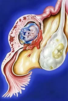
Background imageOviduct Collection: Ectopic pregnancy, artwork C016 / 8737

Ectopic pregnancy, artwork C016 / 8737
Ectopic pregnancy, artwork. This is a pregnancy where the embryo (upper left) implants outside the uterus, usually as here in the Fallopian tube that should channel the fertilised egg to the uterus

Background imageOviduct Collection: Ovulation and fertilisation, artwork

Ovulation and fertilisation, artwork
Ovulation and fertilisation. Computer artwork of the female reproductive system, showing the development of an ovum (egg, small circle) inside an ovary (oval)

Background imageOviduct Collection: Paratubal cysts, light micrograph C015 / 6410

Paratubal cysts, light micrograph C015 / 6410
Paratubal cysts. Light micrograph of a section through a fallopian tube (oviduct) with three paratubal cysts (top). Cysts are fluid-filled sacs

Background imageOviduct Collection: Swollen fallopian tube C015 / 6401

Swollen fallopian tube C015 / 6401
Swollen fallopian tube. This is a condition known as hydrosalpinx, the accumulation of fluid in, and dilation of, a damaged fallopian tube

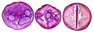
Background imageOviduct Collection: Nematode worm, transverse sections

Nematode worm, transverse sections
Nematode worm. Light micrograph of three transverse sections through the body of a female round unsegmented worm (Ascaris lumbricoides)

Background imageOviduct Collection: Cubic close-packed crystal structure

Cubic close-packed crystal structure
Haemoglobin molecule. Computer artwork showing the structure of a haemoglobin molecule. Haemoglobin is a metalloprotein that transports oxygen around the body in red blood cells

Background imageOviduct Collection: Ovulation, artwork

Ovulation, artwork
Ovulation. Computer artwork of an egg (yellow) being released from an ovary (bottom), a process known as ovulation. The egg is being released into a fallopian tube (top)

Background imageOviduct Collection: False-colour SEM of fimbriae in an embryo

False-colour SEM of fimbriae in an embryo
Fallopian tube in an embryo. False-colour scanning electron micrograph (SEM) of the fimbriae in a ten week old embryo. The fimbriae are the terminal part of the Fallopian tube

Background imageOviduct Collection: Embryo in a fallopian tube, artwork

Embryo in a fallopian tube, artwork
Embryo inside a fallopian tube. Computer artwork representing an embryo (yellow) in the early stages of development inside a fallopian tube. Fallopian tubes lead from the ovaries to the uterus

Background imageOviduct Collection: Fallopian tube

Fallopian tube. Coloured scanning electron micrograph (SEM) of the epithelium of a fallopian tube (oviduct), one of two muscular tubes connecting the ovary to the uterus (womb)

Background imageOviduct Collection: Fimbriae of a Fallopian tube, SEM

Fimbriae of a Fallopian tube, SEM
Fimbriae of a Fallopian tube, coloured scanning electron micrograph (SEM). The fimbriae are folds at the opening of a Fallopian tube, where it opens next to an ovary

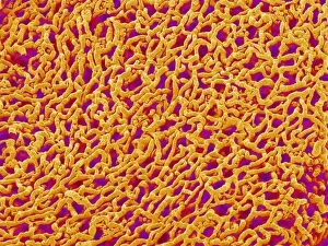
Background imageOviduct Collection: Coloured SEM of fallopian tube cilia & microvilli

Coloured SEM of fallopian tube cilia & microvilli
Fallopian tube cells. Coloured high resolution scanning electron micrograph of cilia and micro- villi on the surface of the fallopian tube epithelium

Background imageOviduct Collection: Fallopian tube blood vessels, SEM

Fallopian tube blood vessels, SEM
Fallopian tube blood vessels. Coloured scanning electron micrograph (SEM) of a cast of blood vessels from a fallopian tube


For sale as Licensed Images

Choose your image, Select your licence and Download the media

The oviduct, also known as the fallopian tube, is a crucial part of the female reproductive system. In this captivating 150-word caption, we delve into the intricate world of female internal organs and explore various aspects related to the oviduct. Starting with a stunning 3D rendering of healthy female internal organs, we witness the beauty and complexity of this delicate system. A comparison between healthy and unhealthy female organs highlights the importance of maintaining optimal reproductive health. Moving closer to the oviduct itself, a light micrograph F006/9799 showcases its intricate structure. Meanwhile, an artwork depicting spider anatomy draws intriguing parallels between different species' reproductive systems. We then shift our focus to fibroid tumors in the uterus, examining their impact on overall reproductive wellness. A conceptual image takes us inside the fallopian tube where sperm meets egg—a pivotal moment in conception. Exploring further, we encounter other essential components such as ovaries and learn about ectopic pregnancy locations. With a three-dimensional view of the entire female reproductive system at hand, we gain a comprehensive understanding of its interconnectedness. Finally, a biomedical illustration demonstrates tubectomy—an important surgical procedure for contraception purposes—ensuring informed choices regarding family planning. This diverse range of visuals provides an insightful journey through various aspects related to oviducts and sheds light on their vital role within female reproduction.