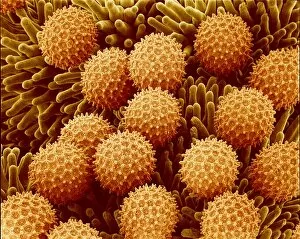
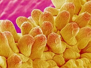
Papilla Collection (#2)

"Papilla: Unveiling the Intricate Surfaces of Nature and Life" Delve into the mesmerizing world of papilla

Background imagePapilla Collection: Tongue papillae, light micrograph

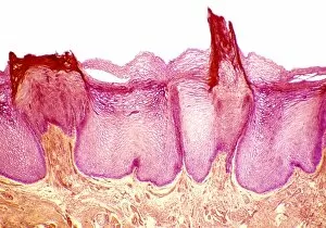
Tongue papillae, light micrograph
Tongue papillae. Coloured light micrograph of a section through the tongue, showing its filiform papillae (red). They project from the tongues upper surface, giving it its rough texture

Background imagePapilla Collection: Taste bud, light micrograph

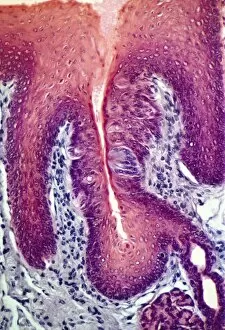
Taste bud, light micrograph
Taste bud. Coloured light micrograph of a section through the tongue, showing a taste bud. This taste bud is seen as a round purple mass slightly to the right of the vertical white line

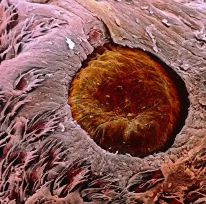
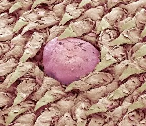
Background imagePapilla Collection: Taste bud, SEM

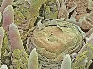
Taste bud, SEM
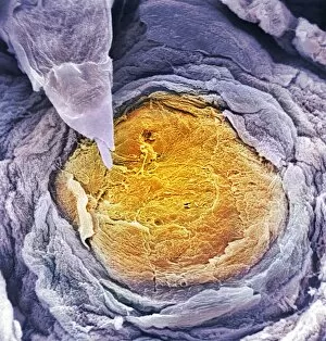
Taste bud. Coloured scanning electron micrograph (SEM) of a taste bud (centre) on a tongue. This is the centre of the round tongue structure called a fungiform papilla

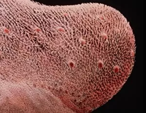
Background imagePapilla Collection: Coloured SEM of a fungiform papilla of the tongue

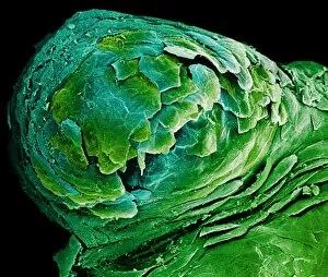
Coloured SEM of a fungiform papilla of the tongue
Fungiform papilla of tongue. Coloured scanning electron micrograph (SEM) of a fungiform papilla of the tongue. The taste buds, the organs of taste, are found under the fungiform papillae

Background imagePapilla Collection: False-colour SEM of fungiform papillae on tongue

False-colour SEM of fungiform papillae on tongue

Background imagePapilla Collection: False-colour SEM of dorsal surface of tongue

False-colour SEM of dorsal surface of tongue
Tongue surface. False-colour scanning electron micrograph (SEM) of the dorsal surface of the tongue. It is covered by two types of projections known as filiform and fungiform papillae

Background imagePapilla Collection: False-colour SEM of a papilla vallata

False-colour SEM of a papilla vallata
Papilla vallata on tongues surface. False-colour scanning electron micrograph of a papilla vallata (brown), a site of taste buds, partly surrounded by filiform papillae (bottom)

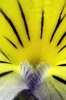
Background imagePapilla Collection: Wild pansy (Viola tricolor) flower

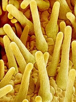
Wild pansy (Viola tricolor) flower. Close-up of a flower from a Viola tricolor plant, showing the papillae (finger-like) and petal structure

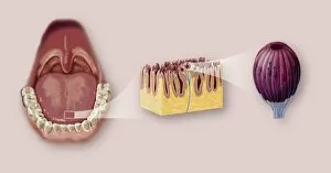
Background imagePapilla Collection: Tongue anatomy, artwork

Tongue anatomy, artwork
Tongue anatomy. Artwork sequence of magnified views of the human mouth and tongue, with the magnification increasing from left to right

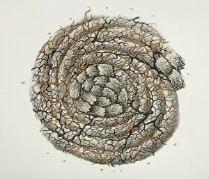
Background imagePapilla Collection: Tongue papilla anatomy, 1844 artwork

Tongue papilla anatomy, 1844 artwork
Tongue papilla anatomy. This anatomical artwork is figure 3, plate 88 bis from volume 3 (1844) of Traite complet de l anatomie de l homme (1831-1854)

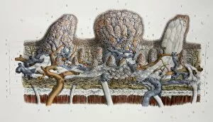
Background imagePapilla Collection: Tongue papillae anatomy, 1844 artwork

Tongue papillae anatomy, 1844 artwork
Tongue papillae anatomy. This anatomical artwork is figure 1, plate 88 from volume 3 (1844) of Traite complet de l anatomie de l homme (1831-1854)

Background imagePapilla Collection: Kudu tongue surface, SEM

Kudu tongue surface, SEM
Kudu tongue surface. Coloured scanning electron micrograph (SEM) of the surface of a kudu (Tragelaphus sp.) antelopes tongue

Background imagePapilla Collection: Squirrel monkey tongue, SEM

Squirrel monkey tongue, SEM
Squirrel monkey tongue. Coloured scanning electron micrograph (SEM) of the surface of a squirrel monkeys (Saimiri sp.) tongue

Background imagePapilla Collection: Cat tongue surface, SEM

Cat tongue surface, SEM
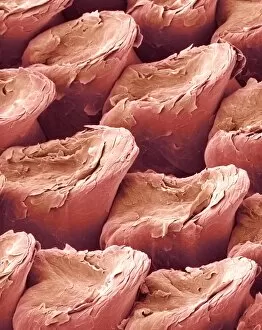
Cats tongue. Coloured scanning electron micrograph (SEM) of the surface of a cats (family Felidae) tongue. The tongue is covered in many backward facing projections called filiform papillae

Background imagePapilla Collection: Human tongue papillae, SEM

Human tongue papillae, SEM
Human tongue papillae. Coloured scanning electron micrograph (SEM) of a fungiform papilla (yellow) and a filiform papilla (spike, top left) from a human tongue

Background imagePapilla Collection: Human tongue surface, SEM

Human tongue surface, SEM
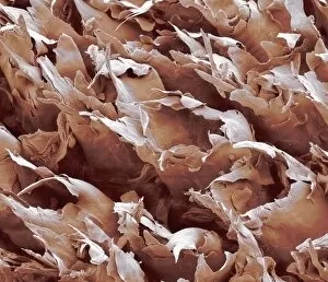
Human tongue surface, coloured scanning electron micrograph (SEM). The tongue is covered in many backward facing projections called filiform papillae, which sense pressure

Background imagePapilla Collection: Tongue surface, SEM

Tongue surface, SEM
Tongue surface, coloured scanning electron micrograph (SEM). The tongue is covered in many backward facing projections called filiform papillae, which sense pressure

Background imagePapilla Collection: Sweet pea flower pollination, SEM

Sweet pea flower pollination, SEM
Sweet pea pollination. Coloured scanning electron micrograph (SEM) of pollen grains (yellow) on the stigma (green) of a sweet pea flower (Lathyrus odoratus)

Background imagePapilla Collection: Morning glory flower pollination, SEM

Morning glory flower pollination, SEM
Morning glory flower pollination. Coloured scanning electron micrograph (SEM) of pollen grains (orange) on the stigma of a morning glory flower (Ipomoea learii)

Background imagePapilla Collection: Comfrey flower petal, SEM

Comfrey flower petal, SEM
Comfrey flower petal. Coloured scanning electron micrograph (SEM) showing papillae on a petal from a comfrey (Symphytum officinale) flower. Papillae are projections from epidermal cells

Background imagePapilla Collection: Flower petal, SEM

Flower petal, SEM
Flower petal. Coloured scanning electron micrograph (SEM) showing papillae (projections) on a petal of a sewer or stink vine flower (Paederia scandens). Magnification: x17 at 6x7cm size

Background imagePapilla Collection: Asian wild flower petal

Asian wild flower petal. Coloured scanning electron micrograph (SEM) of the papillae on the surface of the tube-like petal of Paederia scandens. Papillae are projections from epidermal cells

Background imagePapilla Collection: Morning glory stigma and pollen, SEM

Morning glory stigma and pollen, SEM
Morning glory stigma and pollen. Coloured scanning electron micrograph (SEM) of the stigma (brown) of a morning glory flower (Ipomoea sp.), with pollen grains (orange)

Background imagePapilla Collection: Chrysanthemum petal, SEM

Chrysanthemum petal, SEM
Chrysanthemum petal. Coloured scanning electron micrograph (SEM) of the surface of a petal of a chrysanthemum flower. The lumpy projections are called papillae

Background imagePapilla Collection: Pea flower pollination, SEM

Pea flower pollination, SEM
Pea flower pollination. Coloured scanning electron micrograph (SEM) of pollen grains (yellow) on the stigma (green) of a garden pea flower (Pisum sativum)

Background imagePapilla Collection: Pansy flower petal, SEM

Pansy flower petal, SEM
Pansy flower petal (Viola sp.). Coloured scanning electron micrograph (SEM) showing papillae on a petal from a pansy flower. Papillae are projections from epidermal cells

Background imagePapilla Collection: Geranium flower stigma, SEM

Geranium flower stigma, SEM
Geranium flower stigma. Coloured scanning electron micrograph (SEM) of the stigma of a geranium flower (Geranium thunbergii)

Background imagePapilla Collection: Periwinkle flower pollination, SEM

Periwinkle flower pollination, SEM
Periwinkle flower pollination. Coloured scanning electron micrograph (SEM) of a grain of pollen (lower left) on the stigma of a periwinkle flower (family Apocynaceae)

Background imagePapilla Collection: Pea flower stigma, SEM

Pea flower stigma, SEM
Pea flower stigma. Coloured scanning electron micrograph (SEM) of the surface of a common or garden pea flower (Pisum sativum) stigma

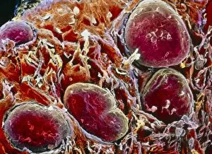
Background imagePapilla Collection: Taste buds, light micrograph

Taste buds, light micrograph
Taste buds. Coloured light micrograph of a section through the tongue, showing taste buds (round, purple). The taste buds are within papillae (projections) located on the surface of the tongue


For sale as Licensed Images

Choose your image, Select your licence and Download the media

"Papilla: Unveiling the Intricate Surfaces of Nature and Life" Delve into the mesmerizing world of papilla, where intricate textures and structures reveal themselves under the lens. From the cat's tongue surface resembling a delicate rasp to forget-me-not flowers showcasing their hidden beauty, these SEM images offer a glimpse into nature's remarkable designs. Intriguingly, even our own bodies hold secrets within. Witness the erect breast nipple captured in an artwork that celebrates human form and diversity. Explore further as we journey beneath the microscope to discover cow tongues' microscopic wonders or delve into cross-sections of a normal heart, unraveling its complex anatomy. But it doesn't stop there; let us guide you through illustrations depicting the structure of human skin and hair. Marvel at how each layer intertwines seamlessly to protect and nourish us from external elements. Delight in diagrams unveiling every detail about this incredible organ that shields our inner selves. And finally, witness feline elegance as we observe cats curling their tongues like ladles to lap up liquid effortlessly. Each papilla pointing backward ensures maximum efficiency while drinking - truly a marvel of evolution. Join us on this captivating journey through papilla's diverse realms - from animal tongues to flower petals, from hearts beating with life to intricate layers protecting our very existence. Prepare for awe-inspiring revelations that will leave you appreciating nature's artistry like never before.