
Papillae Collection (#2)
"Papillae: Unveiling the Intricate World of Texture and Taste" Discover the fascinating realm of papillae, as we delve into their diverse forms and functions


For sale as Licensed Images
Choose your image, Select your licence and Download the media
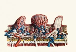
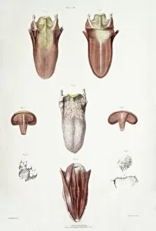
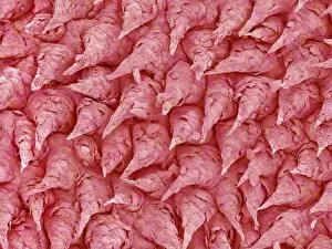
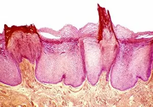
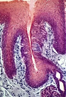
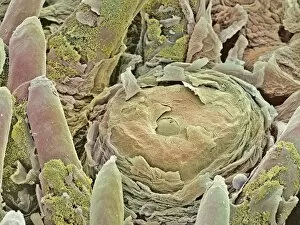
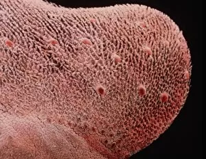
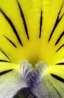
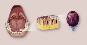
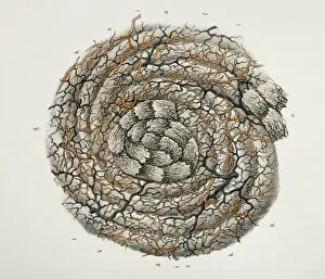
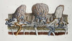
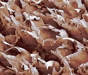
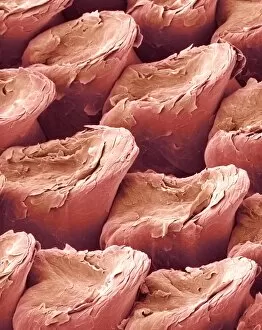
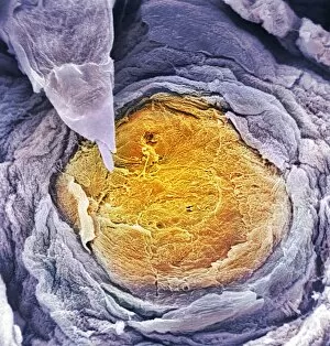
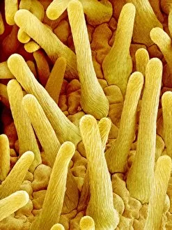
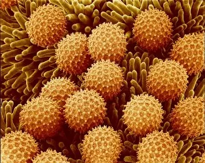
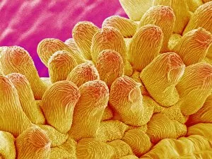
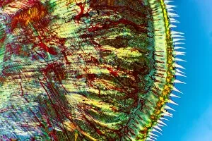
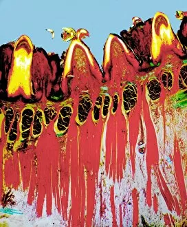
"Papillae: Unveiling the Intricate World of Texture and Taste" Discover the fascinating realm of papillae, as we delve into their diverse forms and functions. These tiny structures, resembling cat tongue surfaces under a scanning electron microscope (SEM), are not only found in feline friends but also play a crucial role in our own gustatory experiences. Intriguingly, SEM images reveal that papillae extend beyond the realms of animals. Take a closer look at the delicate petals of forget-me-not flowers or yarrow flowers, magnified to showcase their intricate textures reminiscent of filiform papilae on our tongues. The digital cross-section illustration further highlights how taste buds intertwine with these unique projections, forming an essential part of our sensory perception. Zooming back into nature's palette, SEM captures mesmerizing details within zinnia flower petals and pansy flower petals. These close-ups unveil an astonishing array of patterns and textures akin to the microscopic world residing on our tongues. But it doesn't stop there. Let's shift gears from flora to fauna as we explore cow tongue under the microscope. Picture No. 11675366 showcases this intriguing organ up close, revealing its own set of distinctive papillae formations that aid bovines in grazing and consuming vegetation effectively. As we immerse ourselves in these captivating visuals (Picture No. 11675345), let us appreciate how something so seemingly insignificant can hold such profound significance for both humans and animals alike. Papillae serve as gatekeepers to flavor sensations; they allow us to savor every delectable bite while ensuring cows graze efficiently on lush pastures. So next time you enjoy your favorite dish or marvel at nature's floral wonders, remember that beneath it all lies a hidden world shaped by intricate papillae – guardians guiding us through tactile exploration and enhancing our connection with food and beauty (Picture No. 11091797, Picture No. 11072772).