Parasitic Collection (#19)

"Exploring the Intricate World Creatures: From Gastrointestinal Nematodes to Eyelash Mites" Delving into the hidden realm of parasites

Background imageParasitic Collection: Downy mildew infection, light micrograph

Downy mildew infection, light micrograph
Downy mildew infection. Light micrograph of a section through a grapevine (Vitis sp.) leaf infected with downy mildew (Plasmopara viticola)

Background imageParasitic Collection: Head of a human flea, light micrograph

Head of a human flea, light micrograph

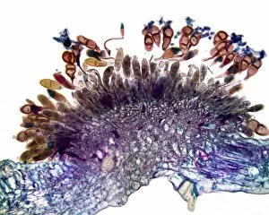
Background imageParasitic Collection: Hollyhock rust fungus in a leaf

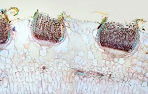
Hollyhock rust fungus in a leaf. Light micrograph of a section through a hollyhock (Alcea sp.) leaf infected with the parasitic Puccinia malvacearum rust, showing the teliospores (spores)

Background imageParasitic Collection: Clubroot infection, light micrograph

Clubroot infection, light micrograph
Clubroot infection. Light micrograph a section through a cabbage root infected with clubroot (Plasmodiophora brassicae). Plasmodiophora is a parasitic protist that infects the roots of cabbages

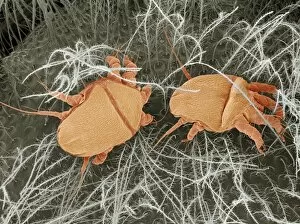
Background imageParasitic Collection: Chicken mite, light micrograph

Chicken mite, light micrograph

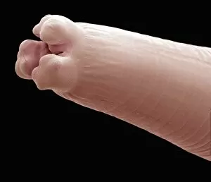
Background imageParasitic Collection: Tapeworm head, SEM

Tapeworm head, SEM
Tapeworm head. Coloured scanning electron micrograph of the head (scolex) of a tapeworm (Taenia pisiformis), showing the hooks used to attach to the host animals digestive tract

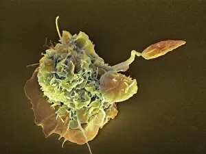
Background imageParasitic Collection: Macrophage attacking a foreign body, SEM

Macrophage attacking a foreign body, SEM
Macrophage attacking a foreign body. Coloured scanning electron micrograph (SEM) of a macrophage white blood cell (round, left) engulfing a Leishmania mexicana protozoan parasite (long, right)

Background imageParasitic Collection: Cistanche phelypaea

Cistanche phelypaea flowering on salt marshes in a Algarve, Portugal. This plant is a parasite on Chenopdiaceae

Background imageParasitic Collection: Teloschistes chrysophthalmus

Teloschistes chrysophthalmus
Golden eye lichen (Teloschistes chrysophthalmus) growing on blackthorn. This is extremely rare in UK, formerly thought to be extinct

Background imageParasitic Collection: Laetiporus sulphureus

Laetiporus sulphureus
Chicken of the Woods (Laetiporus sulphureus) growing on an oak (Quercus) tree

Background imageParasitic Collection: Cytinus hypocistis

Cytinus hypocistis flowering in Algarve, Portugal. This is an ant pollinated root parasite of Cistus species

Background imageParasitic Collection: Naked Broomrape (Orobanche uniflora)

Naked Broomrape (Orobanche uniflora), also known as One-flowered Broomrape, flowering in Waterton, Canada. This plant is parasitic on Round-leaved Alumroot

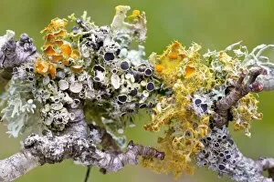
Background imageParasitic Collection: Lichens on Blackthorn

Lichens on Blackthorn
Golden eye lichen (Teloschistes chrysophthalmus) and Physcias growing on blackthorn in unpolluted, humid conditions in Brittany, France

Background imageParasitic Collection: Hoof fungus (Fomitopsis pinicola)

Hoof fungus (Fomitopsis pinicola)
A hoof fungus (Fomitopsis pinicola) growing on a tree stump in Georgia, USA

Background imageParasitic Collection: Malaria life-cycle, artwork

Malaria life-cycle, artwork
Malaria life-cycle. Computer artwork showing the life-cycle and transmission of malaria parasites. Anticlockwise from upper left: A mosquito bites and infects a person

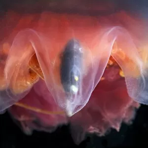
Background imageParasitic Collection: Amphipods inside a hydromedusa

Amphipods inside a hydromedusa. Small crustaceans (Hyperia galba) inside the domed body of a hydromedusa (Sarsia tubulosa). Hydromedusae are the sexually reproductive stage of hydrozoan cnidarians

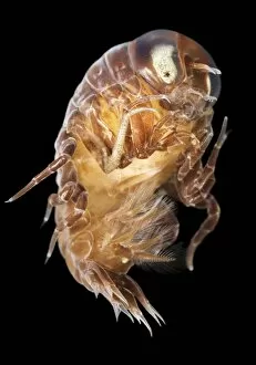
Background imageParasitic Collection: Amphipod

Amphipod. This small crustacean (Hyperia galba) is a parasite of jellyfish. Photographed in the White Sea, Russia

Background imageParasitic Collection: Amphipods inside a moon jellyfish

Amphipods inside a moon jellyfish
Amphipods inside a jellyfish. Small crustaceans (Hyperia galba) inside the domed body of a moon jellyfish (Aurelia aurita)

Background imageParasitic Collection: Amphipod inside a moon jellyfish

Amphipod inside a moon jellyfish
Amphipod inside a jellyfish. Small crustacean (Hyperia galba) inside the domed body of a moon jellyfish (Aurelia aurita). This jellyfish, also known as the common jellyfish

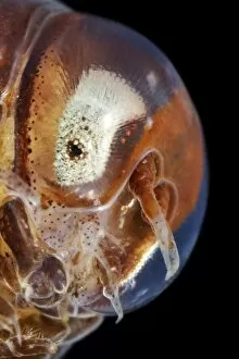
Background imageParasitic Collection: Amphipod head

Amphipod head. This small crustacean (Hyperia galba) is a parasite of jellyfish. Photographed in the White Sea, Russia

Background imageParasitic Collection: Monitor lizard tick

Monitor lizard tick (Amblyomma exornatum, formerly Aponomma exornatum). This tick is native to Africa, where, as the name suggests, it is mainly a parasite of monitor lizards

Background imageParasitic Collection: Seal louse, SEM

Seal louse, SEM
Seal louse (Echinophthirius horridus), coloured scanning electron micrograph (SEM). This ectoparasite (external parasite) feeds on the blood of seals, but only when the seals are on land

Background imageParasitic Collection: Hyobanche sanguinea

Hyobanche sanguinea, a parasitic plant, growing in Namaqualand, South Africa

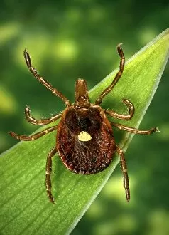
Background imageParasitic Collection: Female lone star tick

Female lone star tick (Amblyomma americanum). This tick is found through the southeast and south-central United States. It is the vector for the spirochete bacteria Borrelia lonestari

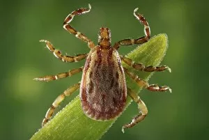
Background imageParasitic Collection: Male yellow dog tick

Male yellow dog tick (Amblyomma aureolatum). This tick is the vector for the bacteria Rickettsia rickettsii in Brazil. Rickettsia rickettsii, causes the often fatal Rocky Mountain spotted fever (RMSF)

Background imageParasitic Collection: Threadworm, SEM

Threadworm, SEM
Threadworm. Coloured scanning electron micrograph (SEM) of the head of a thread worm (Enterobius sp.). This is a nematode worm that is a parasite of the small intestines of numerous animals

Background imageParasitic Collection: Cottony cushion scale bugs

Cottony cushion scale bugs. Cluster of cottony cushion scale (Icerya purchasi) bugs and their egg cases on a lemon tree. Photographed in Italy

Background imageParasitic Collection: Coloured Mistletoe (Viscum coloratum)

Coloured Mistletoe (Viscum coloratum) berries

Background imageParasitic Collection: Malaria infection, artwork

Malaria infection, artwork
Malaria infection. Computer artwork of a macrophage white blood cell (right) extending pseudopodia towards a red blood cell (left) infected with the Plasmodium fulciparum malaria parasite

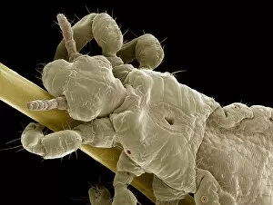
Background imageParasitic Collection: Head louse, SEM

Head louse, SEM
Head louse. Coloured scanning electron micrograph (SEM) of a head louse (Pediculus humanus capitis) clinging to a human hair. Head lice measure 2-3 millimetres in length

Background imageParasitic Collection: Bedbug, SEM

Bedbug, SEM
Bedbug. Coloured scanning electron micrograph (SEM) of the underside of a bedbug (Cimex sp.). Adult bedbugs bodies are flattened, oval, and wingless

Background imageParasitic Collection: Honey bee mites, SEM

Honey bee mites, SEM
Honey bee mites. Coloured scanning electron micrograph (SEM) of bee mites (Varroa sp.) on the body of a bee. Varroa are parasitic mites that attack honey bees (Apis sp.)

Background imageParasitic Collection: Bedbug head, SEM

Bedbug head, SEM
Bedbug head. Coloured scanning electron micrograph (SEM) of the head of a bedbug (Cimex sp.). Antennae are seen at top, with compound eyes (purple) at the sides of its head

Background imageParasitic Collection: Bracket fungus (Laetiporus sulphureus)

Bracket fungus (Laetiporus sulphureus)
Bracket fungus. Young sulphur-coloured fruiting body of the edible bracket fungus known as the chicken of the woods (Laetiporus sulphureus), growing on a living willow tree

Background imageParasitic Collection: Bracket fungus (Daedaleopsis confragosa)

Bracket fungus (Daedaleopsis confragosa)
Blushing bracket fungus (Daedaleopsis confragosa). Close-up of the underside of the blushing bracket fungus showing its pores. The pores are the ends of the tubes within which the spores are produced

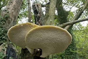
Background imageParasitic Collection: Bracket fungus (Polyporus squamosus)

Bracket fungus (Polyporus squamosus)
Bracket fungus. Dryads saddle (Polyporus squamosus) bracket fungus on a tree

Background imageParasitic Collection: Orobanche minor and Trifolium

Orobanche minor and Trifolium
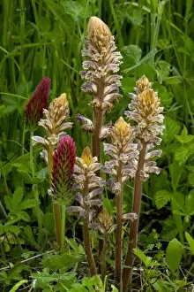
Flowers of Common Broomrape (Orobanche minor) with its host plant, Crimson Clover (Trifolium incarnatum). Photographed in France

Background imageParasitic Collection: Lathraea clandestina

Lathraea clandestina
Flowers of Purple toothwort (Lathraea clandestina) which is a parasitic on Poplars and other trees. Photographed in the Spring in France

Background imageParasitic Collection: Thyme Broomrape (Orobanche alba)

Thyme Broomrape (Orobanche alba) flowers, a parasitic plant on the dead nettle family. Photographed in Greece

Background imageParasitic Collection: Alder bead galls

Alder bead galls on a black alder (Alnus glutinosa) leaf. These galls are caused by the alder gall mite (Phytoptus laevis)

Background imageParasitic Collection: Aphid galls

Aphid galls
Apid galls on a Lombardy poplar (Populus nigra Italica) leaf. The two galls are caused by two different species of aphid. The gall on the leaf stalk is caused by Pemphigus spyrothecae

Background imageParasitic Collection: Pinedrops (Pterospora andromedea )

Pinedrops (Pterospora andromedea )
Pinedrop (Pterospora andromedea ) flowers on a pine forest floor. Photographed in Oregon, USA

Background imageParasitic Collection: Head and thorax of a cat flea, SEM

Head and thorax of a cat flea, SEM
Head and thorax (upper body) of a cat flea (Ctenocephalides felis), coloured scanning electron micrograph (SEM). The head (upper right)

Background imageParasitic Collection: Gall on a yew bud

Gall on a yew bud
Artichoke gall on a yew bud (Taxus sp.). The rounded structure on the end of a branch (lower right) is caused by a gall fly (Taxomyia taxi). Photographed in England

Background imageParasitic Collection: Leaf gall on alder leaves

Leaf gall on alder leaves
Leaf gall (light green spots) on the leaves of an alder tree (Alnus sp.). This leaf damage has been caused by the mite Eriophyes laevis. Photographed in Dorset, England

Background imageParasitic Collection: Rust fungus infection, light micrograph

Rust fungus infection, light micrograph. Transverse section through a barberry (Berberis sp.) leaf infected by a parasitic rust (Puccinia sp.)

Background imageParasitic Collection: Pinedrop (Pterospora andromedea )

Pinedrop (Pterospora andromedea ) flowers. Photographed in Oregon, USA

Background imageParasitic Collection: Toothwort (Lathraea squamaria)

Toothwort (Lathraea squamaria) flowers. This plant is parasitic on the roots of trees including hazel (Corylus sp.), alder (Alnus sp.) and maple (Acer sp.)


For sale as Licensed Images

Choose your image, Select your licence and Download the media

"Exploring the Intricate World Creatures: From Gastrointestinal Nematodes to Eyelash Mites" Delving into the hidden realm of parasites, we encounter gastrointestinal nematodes, microscopic worms that reside in the intestines of various animals. Behold the intricate head structure of a dog tapeworm under scanning electron microscopy (SEM), revealing its remarkable adaptation for survival within its host. Zooming in even closer, SEM unveils the minuscule world of a head louse, showcasing its gripping claws and needle-like mouthparts used for feeding on human blood. Meet the sheep tick, an arachnid parasite found in grassy landscapes; SEM allows us to appreciate its formidable appendages designed for clinging onto hosts like sheep or deer. In our own eyelashes lies an unexpected inhabitant – the eyelash mite. SEM captures their tiny bodies and long tails as they navigate through this often overlooked ecosystem. Journeying deep into Sri Lanka's lush jungles reveals a diverse array organisms coexisting with their hosts amidst nature's beauty and complexity. Witnessing a female mosquito's internal anatomy while sucking blood from human skin through cross-section imagery sheds light on these pesky yet fascinating disease vectors. An illustrated depiction showcases the life cycle and morphology of tapeworms, highlighting their ability to adapt across different host species throughout their complex lifecycle. Examining eyelash mite tails under SEM unravels further mysteries about these elusive creatures that call our lashes home – truly captivating microcosms within ourselves. Venturing underwater brings us face-to-face with sea lampreys; whether it be Petromyzon marinus or other species like lamperns and silver lampreys - these jawless fish are notorious parasites known for attaching themselves to larger marine animals.