
Parasitic Collection (#8)
"Exploring the Intricate World Creatures: From Gastrointestinal Nematodes to Eyelash Mites" Delving into the hidden realm of parasites


For sale as Licensed Images
Choose your image, Select your licence and Download the media
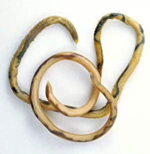
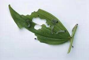
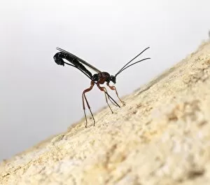
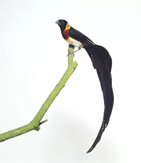
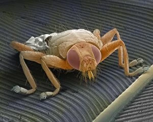
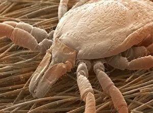
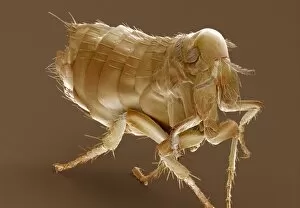
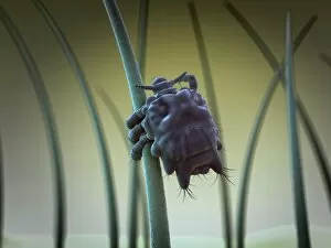
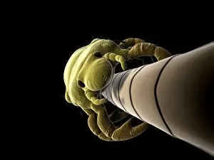
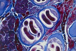
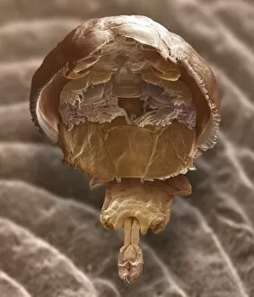
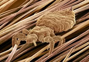
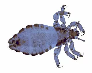
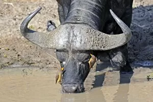
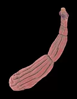
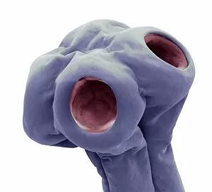
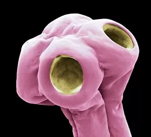
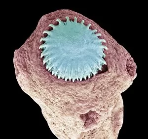
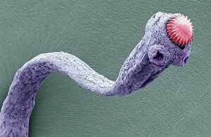
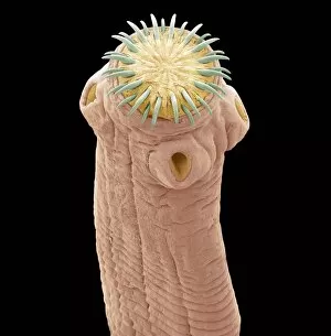
"Exploring the Intricate World Creatures: From Gastrointestinal Nematodes to Eyelash Mites" Delving into the hidden realm of parasites, we encounter gastrointestinal nematodes, microscopic worms that reside in the intestines of various animals. Behold the intricate head structure of a dog tapeworm under scanning electron microscopy (SEM), revealing its remarkable adaptation for survival within its host. Zooming in even closer, SEM unveils the minuscule world of a head louse, showcasing its gripping claws and needle-like mouthparts used for feeding on human blood. Meet the sheep tick, an arachnid parasite found in grassy landscapes; SEM allows us to appreciate its formidable appendages designed for clinging onto hosts like sheep or deer. In our own eyelashes lies an unexpected inhabitant – the eyelash mite. SEM captures their tiny bodies and long tails as they navigate through this often overlooked ecosystem. Journeying deep into Sri Lanka's lush jungles reveals a diverse array organisms coexisting with their hosts amidst nature's beauty and complexity. Witnessing a female mosquito's internal anatomy while sucking blood from human skin through cross-section imagery sheds light on these pesky yet fascinating disease vectors. An illustrated depiction showcases the life cycle and morphology of tapeworms, highlighting their ability to adapt across different host species throughout their complex lifecycle. Examining eyelash mite tails under SEM unravels further mysteries about these elusive creatures that call our lashes home – truly captivating microcosms within ourselves. Venturing underwater brings us face-to-face with sea lampreys; whether it be Petromyzon marinus or other species like lamperns and silver lampreys - these jawless fish are notorious parasites known for attaching themselves to larger marine animals.