Patella Collection (#5)

The patella, also known as the kneecap, is a vital component of the human knee joint and can be seen clearly in a coloured X-ray of a human knee joint

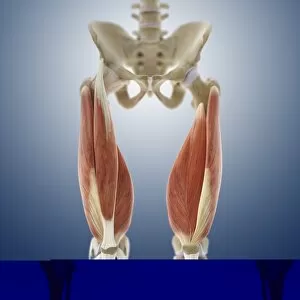
Background imagePatella Collection: Thigh muscles, artwork C013 / 4568

Thigh muscles, artwork C013 / 4568
Thigh muscles. Computer artwork of an anterior view of some of the muscles and tendons (red and white) of the thighs and their attachment to the bones of the leg

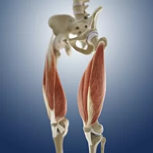
Background imagePatella Collection: Thigh muscles, artwork C013 / 4569

Thigh muscles, artwork C013 / 4569
Thigh muscles. Computer artwork of an oblique anterior view of some of the muscles and tendons (red and white) of the thighs and their attachment to the bones of the leg

Background imagePatella Collection: Human (Homo sapiens) Femur and Patella

Human (Homo sapiens) Femur and Patella
The ball joint from the top end of a human femur and a patella (knee cap) which once belonged to a victim of the Mount Vesuvius eruption of AD 79 which buried the towns of Herculaneum and Pompeii

Background imagePatella Collection: The ' common limpet', is an edible species of sea snail with gills, a typical true limpet

The " common limpet", is an edible species of sea snail with gills, a typical true limpet, a marine gastropod mollusc in the family Patellidae

Background imagePatella Collection: Kneecaps, X-ray

Kneecaps, X-ray
Kneecaps. Coloured X-ray of the kneecaps of a healthy adult man, as viewed from above when the knees are bent. The kneecap (or patella)

Background imagePatella Collection: Healthy knee, X-ray

Healthy knee, X-ray
Knee joint. Coloured X-ray of the healthy knee joint of an adult woman, seen from the side. The knee is the largest joint in the body

Background imagePatella Collection: Knee joint, X-ray

Knee joint, X-ray
Knee joint. Coloured X-ray of the healthy knee joint of a 15-year-old girl. The knee joint is composed of three bones; the femur (thigh bone, upper frame), the tibia (shin bone)

Background imagePatella Collection: Knee replacement, artwork

Knee replacement, artwork
Knee replacement. Artwork of a profile view of a prosthetic knee joint. The prostheses are implanted into the thigh bone (femur) and shinbone (tibia)

Background imagePatella Collection: Kneecap fracture, X-ray

Kneecap fracture, X-ray
Kneecap fracture. Coloured X-ray of a broken kneecap (patella), as viewed from the side with the knee bent. The kneecap (far left, centre) is a disc of bone that covers the front of the knee joint

Background imagePatella Collection: Knee joint from front and side, X-ray

Knee joint from front and side, X-ray
Knee joint from the front (left) and side (right), coloured X-rays. The knee is the largest joint in the human body. It is formed from the articulation of the thigh bone (femur)

Background imagePatella Collection: Artwork of human knee joint, side view

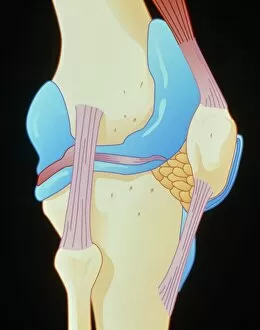
Artwork of human knee joint, side view

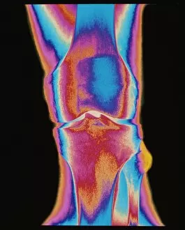
Background imagePatella Collection: Coloured X-ray of a human knee joint (front view)

Coloured X-ray of a human knee joint (front view)
Knee joint. Coloured X-ray of a healthy human knee joint, in front view. Two bones meet at the knee, forming a joint that works like a hinge

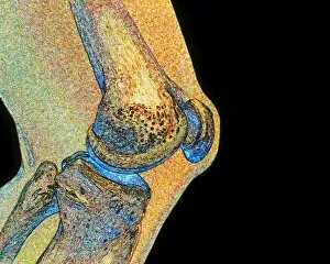
Background imagePatella Collection: Coloured MRI scan of human knee joint, side view

Coloured MRI scan of human knee joint, side view
Knee joint. Coloured Magnetic Resonance Imaging (MRI) scan of a sagittal section through a human knee joint. Two bones (coloured blue) meet at the knee, forming a hinge-joint

Background imagePatella Collection: Coloured X-ray of a human knee joint (side view)

Coloured X-ray of a human knee joint (side view)
Knee joint. Coloured X-ray of a healthy human knee joint in side view, seen while bent. Two bones meet at the knee, forming a joint that works like a hinge

Background imagePatella Collection: Artwork showing the structure of the knee joint

Artwork showing the structure of the knee joint which articulates the femur (upper centre) with the tibia and fibula (bottom)

Background imagePatella Collection: Artwork of bones & ligaments in human knee joint

Artwork of bones & ligaments in human knee joint
Knee joint. Illustration showing bones and tendons of the human knee (genual) joint. The knee is the largest joint in the human body

Background imagePatella Collection: Artwork of bones & ligament in human knee joint

Artwork of bones & ligament in human knee joint
Artwork featuring bones and tendons of the human knee (genual) joint. The single bone of the upper leg (the femur) meets the two smaller bones of the lower limb (the tibia and fibula)

Background imagePatella Collection: Bones of the leg

Bones of the leg. Artwork of human femurs (thigh bones) and patellae (kneecaps), taken from anatomist William Cheseldens textbook Osteographia, or the Anatomy of the Bones, published in 1733

Background imagePatella Collection: Knee bones and ligaments

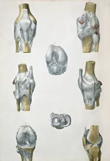
Knee bones and ligaments. Historical anatomical artwork of knee bones (yellow) and ligaments (pale blue). Ligaments are bands of fibrous tissue that hold bones together at joints

Background imagePatella Collection: Arthritis of the knee, X-ray

Arthritis of the knee, X-ray

Background imagePatella Collection: Kneecap fractures, X-ray

Kneecap fractures, X-ray
Kneecap fractures. Coloured X-ray of the kneecap (patella bone) of a 35 year old man, as viewed from above when the knee is bent, showing hairline fractures

Background imagePatella Collection: Knee effusion

Knee effusion. Coloured X-ray of the swelling of an injured knee caused by effusion. Effusion is a painful condition whereby fluid accumulates in the joint space, limiting movement

Background imagePatella Collection: Pinned kneecap fracture, X-ray

Pinned kneecap fracture, X-ray
Pinned kneecap fracture. Coloured X-ray of a pin (grey, centre right) in the fractured kneecap (patella) of an adult man. The metal pin is used to hold the broken bone in the correct position

Background imagePatella Collection: Broken knee, X-ray

Broken knee, X-ray
Broken knee. Coloured X-ray of the knee of a 38 year old patient with a fractured patella (kneecap)

Background imagePatella Collection: Inflamed knee cartilage, computer artwork

Inflamed knee cartilage, computer artwork
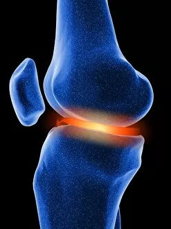
Inflamed knee cartilage. Computer artwork of a knee joint, showing the menisci, cartilage discs that lie between the bones providing a smooth surface for movement

Background imagePatella Collection: Arthrosis of the knee, X-ray

Arthrosis of the knee, X-ray
Arthrosis of the knee, coloured frontal X-ray. The joint is swollen and there are osteophytes, abnormal bony growths (pointed protrusions), seen on the bone at centre

Background imagePatella Collection: Leg bones, artwork

Leg bones, artwork

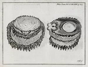
Background imagePatella Collection: Limpet anatomy, 18th century

Limpet anatomy, 18th century

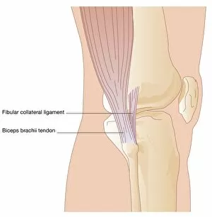
Background imagePatella Collection: Knee in lateral view, artwork

Knee in lateral view, artwork
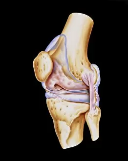
Artwork of the anatomy of the knee joint (lateral view) showing the femur (thigh bone, at top) articulating with the tibia (shin bone, at bottom)

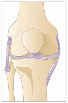
Background imagePatella Collection: Knee in anterior view, artwork

Knee in anterior view, artwork
Artwork of the anatomy of the knee joint (anterior view) showing the femur (thigh bone, at top) articulating with the tibia (shin bone, at bottom), cartilage (light blue)

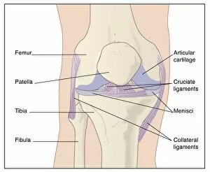
Background imagePatella Collection: Knee joint anatomy, artwork

Knee joint anatomy, artwork
Artwork of the anatomy of the knee joint (anterior view) showing the femur (thigh bone, at top) articulating with the tibia (shin bone, at bottom), articular cartilage

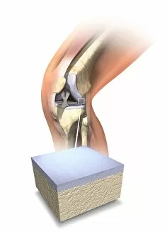
Background imagePatella Collection: Normal human knee, artwork

Normal human knee, artwork
Normal human knee. Artwork showing the internal anatomy of a healthy human knee, with an inset (bottom) of the cartilage (grey) that covers the ends of the bones forming this joint

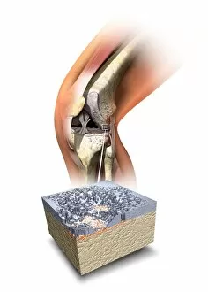
Background imagePatella Collection: Damaged knee, artwork

Damaged knee, artwork
Damaged knee. Artwork showing the internal anatomy of a damaged human knee, with an inset (bottom) of the cartilage (grey) that covers the ends of the bones forming this joint

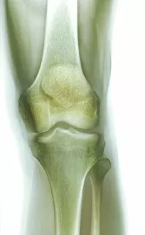
Background imagePatella Collection: Normal knee, X-ray

Normal knee, X-ray
Normal knee, coloured frontal X-ray. The tibia (shin bone, bottom centre) and the smaller fibula (bottom right) articulate with the femur (thigh bone, top) to form the knee joint


For sale as Licensed Images

Choose your image, Select your licence and Download the media

The patella, also known as the kneecap, is a vital component of the human knee joint and can be seen clearly in a coloured X-ray of a human knee joint, standing out amidst the surrounding bones. In a diagram depicting the bones of the right leg and hip, the patella is highlighted as an essential part of this intricate system. When it comes to medical procedures involving the patella, precision is key. A bovie instrument is used to cut through retincaculum and clean up femur in cases where there is displacement or damage to this crucial bone within the knee joint. An illustration showcasing the anterior view of a knee provides insight into its articular surface meniscus. This detailed depiction allows us to understand how these structures work together for optimal movement and stability. Interestingly enough, even nature has its own version of "patella. " In United Kingdom's Scotland region lies Moray's Lossiemouth - home to a vibrant limpet colony on rocks. The blue-rayed limpet (Patella pellucida) thrives here, showcasing its unique adaptation that resembles our very own kneecap. Zooming out from specific anatomy studies, we can appreciate how integral the patella is within our entire skeletal system. Whether viewed from front or side angles with labels or depicted in perspective view alongside other male skeleton parts – it becomes evident that every bone plays an important role in maintaining our body's structure and function. To further highlight diversity across species, one might explore fossils like Mososaurus skull and fish palates. These remnants remind us that while humans have evolved complex anatomical systems such as our knees' intricate design including patellas – other creatures have their own fascinating adaptations too. Whether we examine it through medical images or delve into natural wonders like limpets or fossils – exploring various aspects related to "patella" brings about awe for both human anatomy and biodiversity.