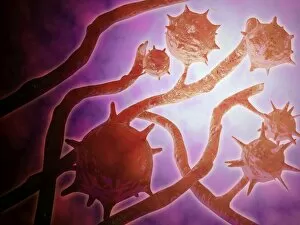
Pathogen Collection (#3)

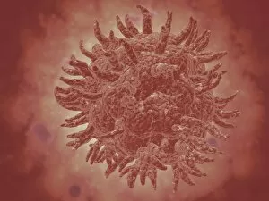
"Unseen Threats: Exploring the World of Pathogens" Budding yeast cell: A microscopic powerhouse, capable of causing infections in humans and animals alike

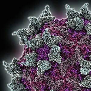
Background imagePathogen Collection: Rhinovirus and antibody, molecular model C015 / 7139

Rhinovirus and antibody, molecular model C015 / 7139
Rhinovirus. Molecular model of the antigen-binding fragment (Fab) from a strongly neutralising antibody bound to a human rhinovirus 14 (HRV-14) particle

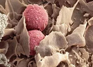
Background imagePathogen Collection: Phagocytosis of fungus spores, SEM

Phagocytosis of fungus spores, SEM
Phagocytosis of fungus spores. Coloured scanning electron micrograph (SEM) of spores from the fungus Aspergillus fumigatus (red) being phagocytosed (engulfed and destroyed) by a dendritic cell (brown)

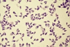
Background imagePathogen Collection: Giardia lamblia protozoa, micrograph

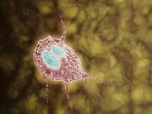
Giardia lamblia protozoa, micrograph
Giardia lamblia protozoa, light micrograph. These single-celled parasitic protozoa infect the intestinal tract and are most common in tropical regions

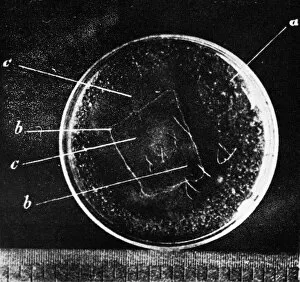
Background imagePathogen Collection: Antibiotic action, historical image

Antibiotic action, historical image
Antibiotic action. Historical image taken by the German pathologist Karl Dohle (1855-1928) in 1889, showing the action of an antibiotic compound on Bacillus anthracis (anthrax)

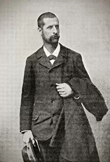
Background imagePathogen Collection: Illustrated portrait of Alexandre Emile Jean Yersin, 1863 - 1943

Illustrated portrait of Alexandre Emile Jean Yersin, 1863 - 1943. Swiss-French physician and bacteriologist. He was the co-discoverer of Yersinia pestis, the bacillus which causes bubonic plague

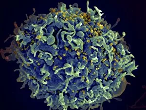
Background imagePathogen Collection: Scanning electron micrograph of HIV particles infecting a human H9 T cell

Scanning electron micrograph of HIV particles infecting a human H9 T cell

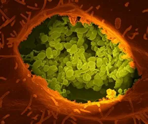
Background imagePathogen Collection: Coxiella burnetii, the bacteria that causes Q Fever

Coxiella burnetii, the bacteria that causes Q Fever. A dry fracture of a Vero cell exposing the contents of a vacuole where Coxiella burnetii are busy growing

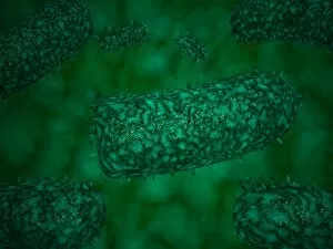
Background imagePathogen Collection: Stylized rabies virus particles

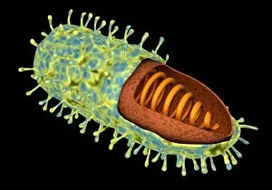
Stylized rabies virus particles, the cause of the viral neuroinvasive disease acute encephalitis

Background imagePathogen Collection: Microscopic view of Neisseria gonorrhoeae

Microscopic view of Neisseria gonorrhoeae. Neisseria gonorrhoeae typically infects the mucous membranes causing infections such as urethritis, cervicitis, salpingitis, pelvic inflammatory disease

Background imagePathogen Collection: Microscopic view of yellow fever virus

Microscopic view of yellow fever virus. Yellow fever is an acute viral disease

Background imagePathogen Collection: Microscopic view of Giardiasis

Microscopic view of Giardiasis, an infectious disease caused by a unicellular parasite known as Giardia lamblia

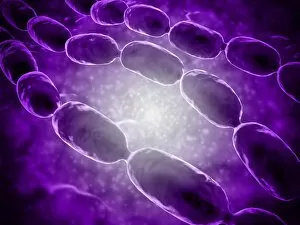
Background imagePathogen Collection: Microscopic view of bacterial pneumonia

Microscopic view of bacterial pneumonia. Bacterial pneumonia is a type of pneumonia caused by bacterial infection. Pneumonia can be generally defined as inflammation of the lung parenchyma

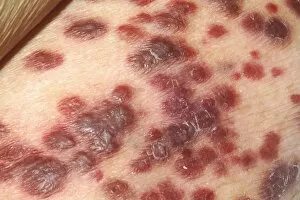
Background imagePathogen Collection: Kaposis sarcoma on the skin of an AIDS patient

Kaposis sarcoma on the skin of an AIDS patient

Background imagePathogen Collection: Microscopic view of Streptococcus pneumoniae

Microscopic view of Streptococcus pneumoniae, also known as pneumococcus. Pneumococcus is a Gram-positive coccus shaped pathogenic bacteria which causes many types of pneumococcal infections in

Background imagePathogen Collection: Microscopic view of Stachybotrys chartarum

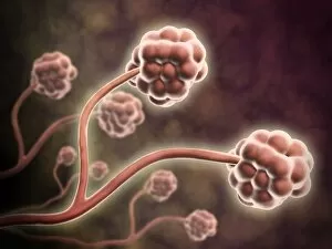
Microscopic view of Stachybotrys chartarum, a black mold

Background imagePathogen Collection: Conceptual image of rabies virus

Conceptual image of rabies virus

Background imagePathogen Collection: HIV infected macrophage, SEM C018 / 8598

HIV infected macrophage, SEM C018 / 8598
HIV infected macrophage. Coloured ion-abrasion scanning electron micrograph (IA-SEM) of a macrophage white blood cell infected with human immunodeficiency virus (HIV, red)

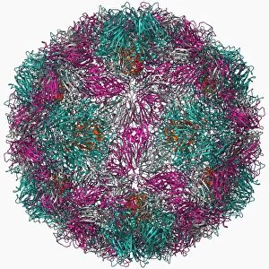
Background imagePathogen Collection: Foot-and-mouth disease virus F006 / 9556

Foot-and-mouth disease virus F006 / 9556
Foot-and-mouth disease virus. Molecular model of the foot-and-mouth disease (FMD) virus (Aphtae epizooticae) protein coat (capsid)

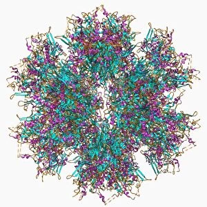
Background imagePathogen Collection: Adenovirus penton base protein F006 / 9542

Adenovirus penton base protein F006 / 9542
Adenovirus penton base protein, molecular model. This protein molecule is a subunit called a penton, forming the vertices of the capsid of this adenovirus

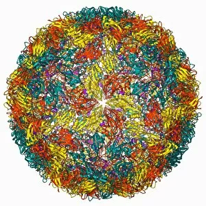
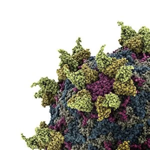
Background imagePathogen Collection: Rhinovirus 16 capsid, molecular model F006 / 9431

Rhinovirus 16 capsid, molecular model F006 / 9431
Rhinovirus 16 capsid, molecular model. This is human rhinovirus 16. The rhinovirus infects the upper respiratory tract and is the cause of the common cold. It is spread by coughs and sneezes

Background imagePathogen Collection: Helicobacter pylori bacteria, SEM C016 / 9136

Helicobacter pylori bacteria, SEM C016 / 9136
Helicobacter pylori bacteria. Coloured scanning electron micrograph (SEM) of Helicobacter pylori bacteria (green) in the stomach. H

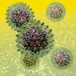
Background imagePathogen Collection: Hepatitis B virus particles, artwork C016 / 9097

Hepatitis B virus particles, artwork C016 / 9097
Hepatitis B virus (HBV) particles, computer artwork. Each particle consists of an outer lipid envelope (green) that surrounds a protein nucleopcapsid (purple)

Background imagePathogen Collection: Rhinovirus and antibody, molecular model C015 / 7138

Rhinovirus and antibody, molecular model C015 / 7138
Rhinovirus. Molecular model of the antigen-binding fragment (Fab) from a strongly neutralising antibody bound to a human rhinovirus 14 (HRV-14) particle

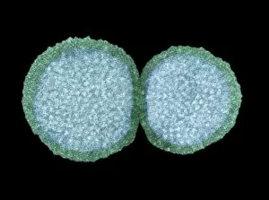
Background imagePathogen Collection: Influenza virus particles, TEM

Influenza virus particles, TEM
Influenza virus particles, coloured transmission electron micrograph (TEM). The virus consists of ribonucleic acid (RNA), surrounded by a nucleocapsid (blue) and a lipid envelope (green)

Background imagePathogen Collection: Propionibacterium acnes C014 / 0631

Propionibacterium acnes C014 / 0631
Propionibacterium acnes bacteria, coloured scanning electron micrograph (SEM). This bacteria is found deep in the hair follicles and pores of the skin

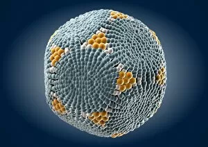
Background imagePathogen Collection: Parvovirus particle, artwork C013 / 4640

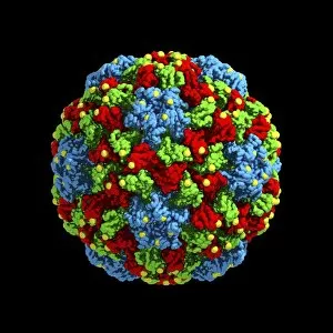
Parvovirus particle, artwork C013 / 4640
Parvovirus particle. Computer artwork showing the outer structure of a human parvovirus (family Parvoviridae) particle (virion)

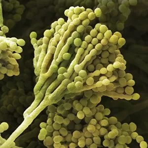
Background imagePathogen Collection: Mould fungus, SEM

Mould fungus, SEM
Mould fungus (Penicillium expansum), coloured scanning electron micrograph (SEM). This fungus, also called blue mould, is a pathogen of plant material

Background imagePathogen Collection: Cucumber mosaic virus, computer model

Cucumber mosaic virus, computer model
Cucumber mosaic virus (CMV), computer model. This image was created using molecular modelling software and data from X-ray crystallography

Background imagePathogen Collection: Murine norovirus with antibody fragments

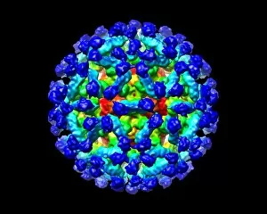
Murine norovirus with antibody fragments
Murine norovirus (MNV) with antibody fragments, computer model. This image was created using molecular modelling software and data from cryo- electron microscopy

Background imagePathogen Collection: AIDS virus

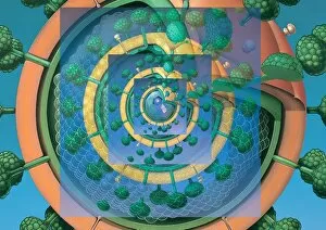
AIDS virus. Conceptual computer artwork of many HIV (human immunodeficiency virus) particles that are sectioned to show their internal structures

Background imagePathogen Collection: Bacteria research

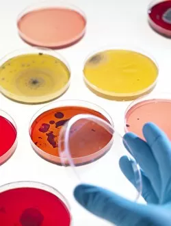
Bacteria research
MODEL RELEASED. Bacteria research. Researcher holding an empty petri dish in front of other dishes containing cultured bacteria (dots)

Background imagePathogen Collection: Smallpox virus particle, TEM

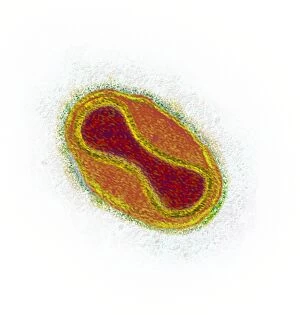
Smallpox virus particle, TEM
Smallpox virus particle. Coloured transmission electron micrograph (TEM) of a Variola major virus particle. Each particle consists of a DNA (deoxyribonucleic acid)

Background imagePathogen Collection: Infectious bronchitis virus (IBV), TEM

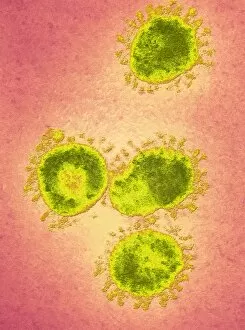
Infectious bronchitis virus (IBV), TEM
Infectious bronchitis virus (IBV) particles, coloured transmission electron micrograph (TEM). This virus is a member of the coronavirus family

Background imagePathogen Collection: Eastern equine encephalitis virus, TEM

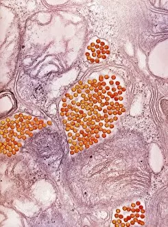
Eastern equine encephalitis virus, TEM
Eastern equine encephalitis virus. Coloured transmission electron micrograph (TEM) of eastern equine encephalitis (EEE) virus particles (Alphavirus sp. orange) in the salivary gland of a mosquito

Background imagePathogen Collection: HIV particles, TEM

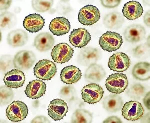
HIV particles, TEM
HIV (human immunodeficiency virus) particles, coloured transmission electron micrograph (TEM). Each particle consists of an RNA (ribonucleic acid, purple/brown) genome

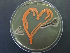
Background imagePathogen Collection: Love, microbial art

Love, microbial art. Petri dish containing the fungi Aureobasidium pullulans and Rhodotorula sp. (red). The fungi have been cultured on agar jelly to depict a heart and arrow

Background imagePathogen Collection: Bacillus subtilis bacteria, SEM

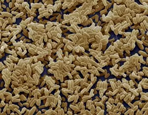
Bacillus subtilis bacteria, SEM
Bacillus subtilis. Coloured scanning electron micrograph (SEM) of Bacillus subtilis bacteria. B. subtilis, or hay bacillus, is an aerobic, gram-positive bacillus(rod-shaped bacterium)

Background imagePathogen Collection: Bacteria on lung hairs

Bacteria on lung hairs
Bacteria in a lung. Computer artwork of bacteria (yellow) on cilia (hair-like structures) in a lung. Bacterial infection of the lungs can cause potentially fatal pneumonia

Background imagePathogen Collection: Spirochete bacteria, TEM

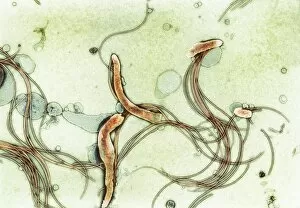
Spirochete bacteria, TEM
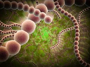
Spirochete bacteria, coloured transmission electron micrograph (TEM). Spirochetes are bacteria with helically coiled cells

Background imagePathogen Collection: Yeast cells, TEM

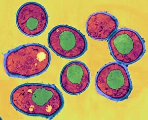
Yeast cells, TEM
Yeast cells, coloured transmission electron micrograph (TEM). The cell wall is shown in blue, cytoplasm in red, and vacuole in green. Yeast is a single-celled fungus that reproduces asexually

Background imagePathogen Collection: Fungal spores, SEM

Fungal spores, SEM
Fungal spores. Coloured scanning electron micrograph (SEM) of fungal spores on a fruiting body of an Aspergillus sp. fungus. The name for such a fruiting body is a conidiophore

Background imagePathogen Collection: Stem rust, Puccinia, wheat, Triticum aestivum

Stem rust, Puccinia, wheat, Triticum aestivum - By Franz Bauer. Paper, watercolour, signed. London, Natural History Museum, Library and Archives, Unfinished Drawings of Plants, vol. 6, no. 77

Background imagePathogen Collection: Illustration of immune response, involving chain of defensive white blood cells, triggered by microb

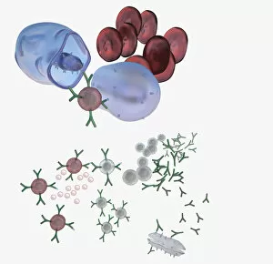
Illustration of immune response, involving chain of defensive white blood cells, triggered by microb

Background imagePathogen Collection: Cross section biomedical illustration on grid of Worm, Fungi, Protozoa

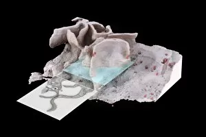
Cross section biomedical illustration on grid of Worm, Fungi, Protozoa, and Bacteria infection and infestation

Background imagePathogen Collection: Single virus particle

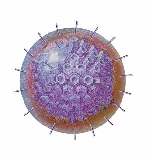
Single virus particle

Background imagePathogen Collection: Red blood cells on an agar plate are used to diagnose infection

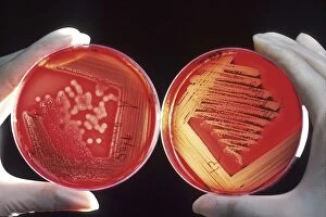
Red blood cells on an agar plate are used to diagnose infection. The plate on the left shows a positive staphyloccus infection

Background imagePathogen Collection: Conceptual image of antibody attaching and killing bacteria

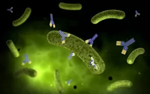
Conceptual image of antibody attaching and killing bacteria

Background imagePathogen Collection: Microscopic view of histoplasmosis

Microscopic view of histoplasmosis. Histoplasmosis is an infection in the lungs caused by inhaling the spores of a fungus. This fungus, called Histoplasma capsulatum


For sale as Licensed Images

Choose your image, Select your licence and Download the media

"Unseen Threats: Exploring the World of Pathogens" Budding yeast cell: A microscopic powerhouse, capable of causing infections in humans and animals alike. Avian flu virus: A notorious pathogen that poses a significant threat to both birds and humans, with potential for devastating global outbreaks. Neutrophil engulfing MRSA, SEM C018 / 8596: Witness the incredible defense mechanism of our immune system as it battles against antibiotic-resistant bacteria. E. Coli bacteria, SEM: Unveiling the minuscule culprits behind foodborne illnesses and urinary tract infections. Salmonella bacteria, SEM: Discover the intricate structure of this common bacterial pathogen responsible for causing severe gastrointestinal distress. Candida fungus, SEM: Peering into the world of fungal infections as Candida takes center stage in various ailments from thrush to systemic candidiasis. Coral Spot Fungus (Nectria cinnabarina) fruiting bodies on Sycamore twig, Powys, Wales: Marvel at nature's ability to harbor pathogens even within picturesque settings like these vibrant red fruiting bodies growing on trees. HIV particle: Delve into the complex realm of retroviruses with a focus on HIV/AIDS – an ongoing global health crisis demanding attention and research breakthroughs. Anthrax cultures, historical diagram: Tracing back through history to understand one of humanity's oldest known pathogens – anthrax – its deadly impact and potential bioweapon applications throughout time. Norovirus particles, TEM: Get up close with these tiny viral particles responsible for stomach flu outbreaks worldwide; their resilience challenges public health measures constantly. Tuberculosis bacteria - Explore the persistent Mycobacterium tuberculosis that causes tuberculosis disease affecting millions globally despite medical advancements Avian flu virus - Investigate avian influenza viruses' ability to jump species barriers, posing a constant threat to both birds and humans.