Pathogen Collection (#48)

"Unseen Threats: Exploring the World of Pathogens" Budding yeast cell: A microscopic powerhouse, capable of causing infections in humans and animals alike

Background imagePathogen Collection: Bacteria

Bacteria. Computer artwork of a multitude of spherical (cocci) bacteria. Many bacteria have this shape, including Streptococcus, which live normally in the human body but can be pathogenic

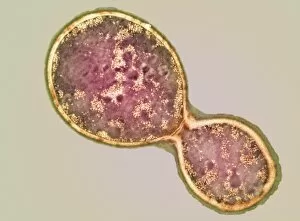
Background imagePathogen Collection: Cryptococcus cell dividing, TEM

Cryptococcus cell dividing, TEM
Cryptococcus cell dividing. Coloured transmission electron micrograph (TEM) of the asexual budding (lower right) of a Cyptococcus sp. yeast cell

Background imagePathogen Collection: Fungus on maple leaf

Fungus on maple leaf. Unidentified fungus (red) growing on the tips of maple (Acer sp.) leaves. Photographed in the Lower Stein Valley, southern British Columbia, Canada

Background imagePathogen Collection: Bleeding canker on tree bark

Bleeding canker on tree bark
Bleeding canker on a horse chestnut tree (Aesculus hippocastanum). Where this pathogen has been active, bark, cambial tissue and the phloem (sugar conducting tissue) are destroyed

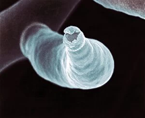
Background imagePathogen Collection: Fungal reproduction, SEM

Fungal reproduction, SEM
Fungal reproduction. Coloured scanning electron micrograph (SEM) of the tip of a branch of a conidiophore (one type of fungal reproductive structure) with a conidia (spore) emerging from the tip

Background imagePathogen Collection: Bleeding canker

Bleeding canker. Liquid exuding from the trunk of a horse chestnut tree (Aesculus hippocastanum). This pathogen destroys bark, cambial tissue and the phloem (sugar conducting tissue)

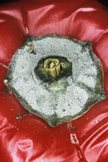
Background imagePathogen Collection: Mouldy pepper

Mouldy pepper. Grey mould (Botrytis cinerea) on a red pepper (Capsicum sp.)

Background imagePathogen Collection: Gut bacterium reproducing, TEM

Gut bacterium reproducing, TEM
Gut bacterium reproducing. Coloured transmission electron micrograph (TEM) of a single bacterium dividing into two identical clone daughter bacteria

Background imagePathogen Collection: Plague bacteria

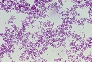
Plague bacteria. Light micrograph of Gram- stained Yersinia pestis bacteria. This bacterium is the cause of bubonic plague (generally agreed to have been the Black Death of the Middle Ages)

Background imagePathogen Collection: Bacteria, computer artwork

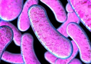
Bacteria, computer artwork
Bacteria. Computer artwork of a multitude of rod-shaped (bacillus) bacteria. Many bacteria have this shape, including Escherichia coli, which can cause food poisoning, and Bacillus anthracis

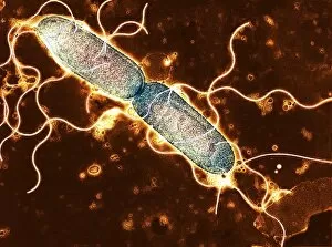
Background imagePathogen Collection: Salmonella, TEM

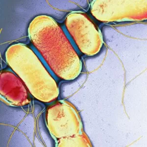
Salmonella, TEM
Salmonella. Coloured transmission electron micrograph (TEM) of Salmonella paratyphi B bacteria (also called Salmonella schotmulleri)

Background imagePathogen Collection: S. maltophilia bacteria, TEM

S. maltophilia bacteria, TEM
Stenotrophomonas maltophilia bacteria, coloured transmission electron micrograph (TEM). This aerobic Gram-negative bacterium, previously known as Pseudomonas maltophilia

Background imagePathogen Collection: Germ, conceptual computer artwork

Germ, conceptual computer artwork

Background imagePathogen Collection: Q fever bacteria, Coxiella burnetii, TEM

Q fever bacteria, Coxiella burnetii, TEM
Q fever bacteria (Coxiella burnetii), coloured transmission electron micrograph (TEM). Q fever is a disease present in livestock that can be spread to humans by inhalation of contaminated particles

Background imagePathogen Collection: Vibrio vulnificus bacteria, SEM

Vibrio vulnificus bacteria, SEM
Vibrio vulnificus bacteria, coloured scanning electron micrograph (SEM). This Gram-negative rod- shaped motile bacteria is found in sea water

Background imagePathogen Collection: Clostridium botulinum bacteria

Clostridium botulinum bacteria, computer artwork. This Gram-positive rod-shaped bacteria causes the disease botulism, a type of food poisoning. The bacteria is found naturally in soil

Background imagePathogen Collection: Clostridium difficile bacteria, TEM

Clostridium difficile bacteria, TEM
Clostridium difficile drug-resistant bacteria, coloured transmission electron micrograph (TEM). This bacterium, also known as C. diff, is a normal member of the intestinal flora in humans

Background imagePathogen Collection: Gonorrhoea bacteria, TEM

Gonorrhoea bacteria, TEM
Gonorrhoea bacteria. Coloured transmission electron micrograph (TEM) of a Neisseria gonorrhoeae bacteria diplococci (pair of cells, light orange) infecting a human epithelial cell (dark orange)

Background imagePathogen Collection: Rod-shaped bacteria, artwork

Rod-shaped bacteria, artwork
Rod-shaped bacteria. Computer artwork of rod-shaped bacteria (bacilli) on the stomach lining. Many bacteria have this shape, including Escherichia coli

Background imagePathogen Collection: Chlamydia pneumoniae bacterium

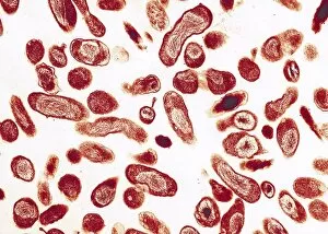
Chlamydia pneumoniae bacterium
Chlamydia pneumoniae. Coloured transmission electron micrograph of a sectioned Chlamydia pneumoniae bacteria. This Gram-negative bacterium is pear-shaped

Background imagePathogen Collection: Proteus vulgaris bacteria, SEM

Proteus vulgaris bacteria, SEM
Proteus vulgaris bacteria, scanning electron micrograph (SEM). The fine threadlike projections are flagella, which allow the bacterium to move. P

Background imagePathogen Collection: Salmonella bacterium, computer artwork

Salmonella bacterium, computer artwork. This is a Gram-negative rod-shaped bacteria that has flagella (hair-like structures) that it uses for locomotion

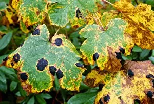
Background imagePathogen Collection: Tar spot fungus on sycamore leaves

Tar spot fungus on sycamore leaves. Leaves of a sycamore (Acer pseudoplatanus) tree, affected by the fungal pathogen Rhytisma acerinum, which causes a disease known as tar spot

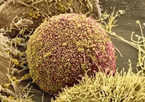
Background imagePathogen Collection: Cell infected with HIV, SEM

Cell infected with HIV, SEM
Cell infected with HIV. Coloured scanning electron micrograph (SEM) of HIV particles (yellow) budding from the membrane of a host cell (red)

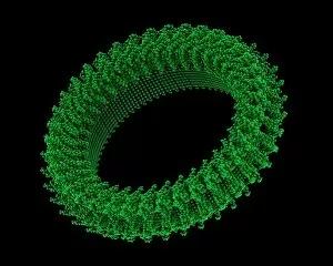
Background imagePathogen Collection: Pore forming bacterial toxin

Pore forming bacterial toxin. Computer model showing the molecular structure of the bacterial toxin pneumolysin. Pneumolysin is a pore forming toxin (PFT)

Background imagePathogen Collection: Linezolid antibiotic, molecular model

Linezolid antibiotic, molecular model
Linezolid antibiotic drug, molecular model. This new drug, marketed as Zyvoxid and Zyvox, is used to treat infections by bacteria, such as MRSA, that are resistant to other antibiotics


For sale as Licensed Images

Choose your image, Select your licence and Download the media

"Unseen Threats: Exploring the World of Pathogens" Budding yeast cell: A microscopic powerhouse, capable of causing infections in humans and animals alike. Avian flu virus: A notorious pathogen that poses a significant threat to both birds and humans, with potential for devastating global outbreaks. Neutrophil engulfing MRSA, SEM C018 / 8596: Witness the incredible defense mechanism of our immune system as it battles against antibiotic-resistant bacteria. E. Coli bacteria, SEM: Unveiling the minuscule culprits behind foodborne illnesses and urinary tract infections. Salmonella bacteria, SEM: Discover the intricate structure of this common bacterial pathogen responsible for causing severe gastrointestinal distress. Candida fungus, SEM: Peering into the world of fungal infections as Candida takes center stage in various ailments from thrush to systemic candidiasis. Coral Spot Fungus (Nectria cinnabarina) fruiting bodies on Sycamore twig, Powys, Wales: Marvel at nature's ability to harbor pathogens even within picturesque settings like these vibrant red fruiting bodies growing on trees. HIV particle: Delve into the complex realm of retroviruses with a focus on HIV/AIDS – an ongoing global health crisis demanding attention and research breakthroughs. Anthrax cultures, historical diagram: Tracing back through history to understand one of humanity's oldest known pathogens – anthrax – its deadly impact and potential bioweapon applications throughout time. Norovirus particles, TEM: Get up close with these tiny viral particles responsible for stomach flu outbreaks worldwide; their resilience challenges public health measures constantly. Tuberculosis bacteria - Explore the persistent Mycobacterium tuberculosis that causes tuberculosis disease affecting millions globally despite medical advancements Avian flu virus - Investigate avian influenza viruses' ability to jump species barriers, posing a constant threat to both birds and humans.