Pathogens Collection (#4)

"Unseen Threats: Exploring the World of Pathogens" Nature's Battle: A tobacco hornworm feasting on a tobacco plant

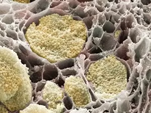
Background imagePathogens Collection: Cabbage root infection, SEM

Cabbage root infection, SEM
Cabbage root infection. Coloured scanning electron micrograph (SEM) of a cross-section through one of the roots of a cabbage (Brassica sp)

Background imagePathogens Collection: Erwinia bacteria, SEM

Erwinia bacteria, SEM
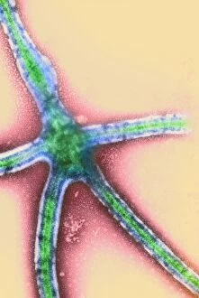
Erwinia bacteria. Coloured scanning electron micrograph (SEM) of Erwinia carotovora bacteria. This gram-negative flagellated bacterium causes soft rot of plant crops such as potato

Background imagePathogens Collection: Cultivated Crabapple (Malus sp. ) close-up of leaves, infected with Cedar-apple Rust

Cultivated Crabapple (Malus sp. ) close-up of leaves, infected with Cedar-apple Rust
Cultivated Crabapple (Malus sp.) close-up of leaves, infected with Cedar-apple Rust (Gymnosporangium juniperi-virginianae), U.S.A

Background imagePathogens Collection: Barley (Hordeum vulgare) close-up of ears, infected with Loose Smut (Ustilago nuda f. sp)

Barley (Hordeum vulgare) close-up of ears, infected with Loose Smut (Ustilago nuda f. sp. hordei) fungal disease
Barley (Hordeum vulgare) close-up of ears, infected with Loose Smut (Ustilago nuda f.sp.hordei) fungal disease, Lincolnshire, England, june

Background imagePathogens Collection: Grasshopper (Orthoptera sp. ) adult, killed by Pathogenic Fungus (Entomophthora sp)

Grasshopper (Orthoptera sp. ) adult, killed by Pathogenic Fungus (Entomophthora sp)
Grasshopper (Orthoptera sp.) adult, killed by Pathogenic Fungus (Entomophthora sp.) infection, fungus causes grasshopper to climb stalk before dying so spores will spread further, Pyrenees, Ariege

Background imagePathogens Collection: Sycamore Tar Spot (Rhytisma acerinum) lesions on fallen Sycamore (Acer pseudoplatanus) leaves

Sycamore Tar Spot (Rhytisma acerinum) lesions on fallen Sycamore (Acer pseudoplatanus) leaves, covered with light snow, Romania, october

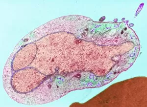
Background imagePathogens Collection: Naegleria fowleri protozoa, TEM

Naegleria fowleri protozoa, TEM
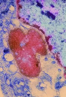
Naegleria fowleri protozoan. Coloured transmission electron micrograph (TEM) of a section through a Naegleria fowleri protozoan

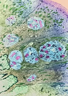
Background imagePathogens Collection: Enterocytozoon sp. parasites, TEM

Enterocytozoon sp. parasites, TEM

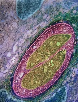
Background imagePathogens Collection: Isospora sp. parasites, TEM

Isospora sp. parasites, TEM
Isospora sp. parasites. Coloured transmission electron micrograph (TEM) of Isospora sp. parasites (yellow) in a sectioned cell

Background imagePathogens Collection: Nanorobots killing bacteria

Nanorobots killing bacteria
Medical nanorobots. Computer artwork of nanorobots killing bacteria (red). These microscopic robots are a future technology that could be developed to help treat diseases

Background imagePathogens Collection: Coxsackie virus particles, TEM

Coxsackie virus particles, TEM
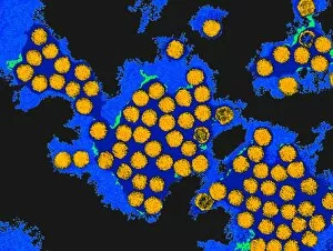
Coxsackie virus particles, coloured transmission electron micrograph (TEM). Coxsackie viruses were named after the town in the USA where they were isolated

Background imagePathogens Collection: Hepatitis A viruses, TEM

Hepatitis A viruses, TEM
Hepatitis A viruses. Coloured transmission electron micrograph (TEM) of hepatitis A viruses, a cause of liver inflammation. Hepatitis A is transmitted through infected food or drink

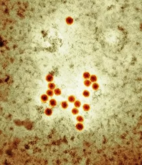
Background imagePathogens Collection: Hepatitis B viruses, TEM

Hepatitis B viruses, TEM
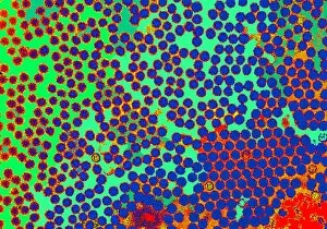
Hepatitis B viruses. Coloured transmission elect- ron micrograph (TEM) of hepatitis B viruses (large lime green circles), a cause of liver inflamm- ation

Background imagePathogens Collection: Virus research, artwork

Virus research, artwork
Virus research. Conceptual computer artwork representing research into viruses, showing a virus particle (lower left) and a computer screen displaying data on the virus

Background imagePathogens Collection: Macrophage engulfing bacteria, SEM

Macrophage engulfing bacteria, SEM
Macrophage engulfing bacteria. Coloured scanning electron micrograph (SEM) of a macrophage white blood cell (brown) engulfing Mycobacterium tuberculosis bacteria (yellow)

Background imagePathogens Collection: Moth virus

Moth virus

Background imagePathogens Collection: Ebola virus replication, TEM

Ebola virus replication, TEM

Background imagePathogens Collection: Dengue fever virus particles, TEM

Dengue fever virus particles, TEM

Background imagePathogens Collection: Dengue fever virus replication, TEM

Dengue fever virus replication, TEM

Background imagePathogens Collection: Malaria parasite, TEM

Malaria parasite, TEM
Malaria parasite. Image 4 of 10. Coloured transmission electron micrograph (TEM) of a sexual male malaria (Plasmodium sp.) microgametocyte in a mosquito (Anopheles sp.) gut

Background imagePathogens Collection: Urinary tract infection, computer artwork

Urinary tract infection, computer artwork
Urinary tract infection. Conceptual computer artwork of bacteria (green) in the urinary tract (represented by metal tubes)

Background imagePathogens Collection: Mosaic plant viruses, TEM

Mosaic plant viruses, TEM
Turnip yellow mosaic viruses. Coloured transmission electron micrograph (TEM) of turnip yellow mosaic viruses (TYMV). This virus infects crucifer plants such as turnip, swede and cauliflower

Background imagePathogens Collection: Influenza virus particles

Influenza virus particles. Computer artwork of influenza (flu) virus particles. Each virus consists of a core of RNA (ribonucleic acid) genetic material surrounded by a protein coat

Background imagePathogens Collection: Influenza viruses

Influenza viruses. Coloured transmission electron micrograph (TEM) of influenza (flu) viruses. The flu virus belongs to the orthomyxovirus group of viruses which have an affinity for mucus

Background imagePathogens Collection: Adenoviruses

Adenoviruses

Background imagePathogens Collection: Virus replication cycle, artwork

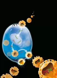
Virus replication cycle, artwork. The cycle starts at upper centre, as virus particles (orange) enter a cell (blue). Once in the cell the viral genetic material, deoxyribonucleic acid (DNA, green)

Background imagePathogens Collection: Budding HIV particle, computer artwork

Budding HIV particle, computer artwork
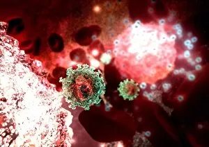
Budding HIV particle. Computer artwork of an HIV (human immunodeficiency virus) particle (green) exiting a T-lymphocyte (a type of white blood cell, lower left)

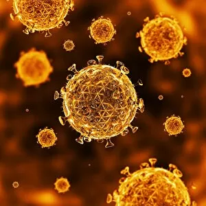
Background imagePathogens Collection: HIV particles

HIV particles, computer artwork. HIV (human immunodeficiency virus) causes AIDS (acquired immune deficiency syndrome). The virus consists of an RNA genome

Background imagePathogens Collection: Simian AIDS viruses, TEM

Simian AIDS viruses, TEM
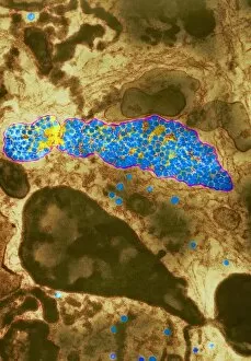
Simian AIDS virus particles, coloured transmission electron micrograph (TEM). These virus particles (small, round) are bursting out of a cell (across bottom) after using the cell to replicate

Background imagePathogens Collection: Vaccinia viruses, TEM

Vaccinia viruses, TEM
Vaccinia virus particles. Coloured transmission electron micrograph (TEM) of sectioned vaccinia virus particles. The genetic cores (red) are covered by membrane layers (green)

Background imagePathogens Collection: Yellow fever virus particles, TEM

Yellow fever virus particles, TEM
Yellow fever virus particles. Coloured transmission electron micrograph (TEM) of yellow fever virus particles (white) growing in culture

Background imagePathogens Collection: Crimean-Congo haemorrhagic fever virus

Crimean-Congo haemorrhagic fever viruses. Coloured transmission electron micrograph (TEM) of Crimean- Congo haemorrhagic fever (CCHF) viruses (brown)

Background imagePathogens Collection: HIV virus particles, artwork

HIV virus particles, artwork
HIV virus particles, computer artwork. Each particle (virion) consists of RNA (ribonucleic acid) contained within a capsid and an envelope covered in glycoprotein spikes


For sale as Licensed Images

Choose your image, Select your licence and Download the media

"Unseen Threats: Exploring the World of Pathogens" Nature's Battle: A tobacco hornworm feasting on a tobacco plant, showcasing the intricate relationship between pathogens and their hosts. Fungal Invasion: Coral Spot Fungus (Nectria cinnabarina) fruiting bodies thriving on a Sycamore twig in Powys, Wales – a reminder of how they are infiltrate even the hardiest organisms. Tiny Terrors: Norovirus particles captured under an electron microscope, revealing the minuscule yet potent nature of these infectious agents. The Silent Killer: Tuberculosis bacteria lurking within our midst, highlighting the ongoing battle against this ancient disease that still plagues humanity today. Unwelcome Guests: Salmonella bacteria magnified through scanning electron microscopy, serving as a stark reminder of foodborne illnesses and their potential consequences. Airborne Hazards: Artwork depicting infections spread by sneezing – emphasizing how easily they are travel and infect others within close proximity. Cellular Warriors: Macrophage engulfing TB bacteria showcased under SEM, illustrating our immune system's relentless fight against invading pathogens. Invisible Invaders: Intestinal protozoan parasites seen through transmission electron microscopy; uncovering the hidden world where these microscopic creatures wreak havoc on human health. Waterborne Nightmare: Cryptosporidium protozoa viewed under TEM – shedding light on water-related diseases caused by these resilient parasites that pose risks to both humans and animals alike. Emerging Perils: Rift Valley fever virus particles revealed through TEM imagery; underscoring the constant threat posed by emerging viruses with potentially devastating consequences for global health security. Mosquito Menace: Dengue fever virus particles visualized using TEM; reminding us of mosquito-borne diseases' impact worldwide and efforts to combat them effectively. The Immune Battle Continues.