Pathology Collection (#3)

"Exploring the Intricate World of Pathology: Unveiling the Mysteries Within" Delving into the realm of pathology, we encounter a fascinating array of scientific wonders

Background imagePathology Collection: Jean-Martin Charcot

Jean-Martin Charcot (1825-1893) - French neurologist and professor of anatomical pathology Date: circa 1890

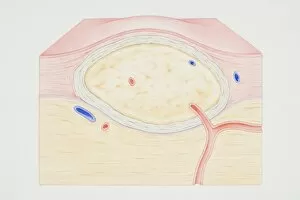
Background imagePathology Collection: Cross-section diagram of a non-cancerous tumour including a fibrous capsule

Cross-section diagram of a non-cancerous tumour including a fibrous capsule, tissue layer and blood vessel

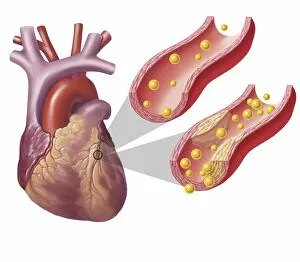
Background imagePathology Collection: Heart with arteries showing cholesterol in one and plaque in the other

Heart with arteries showing cholesterol in one and plaque in the other
Heart with arteries showing cholesterol in one artery and atherosclerotic plaque in the other

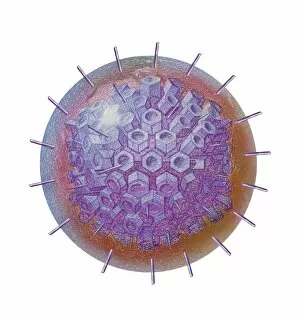
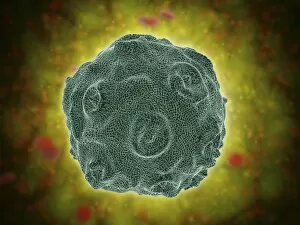
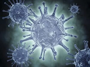
Background imagePathology Collection: Single virus particle

Single virus particle

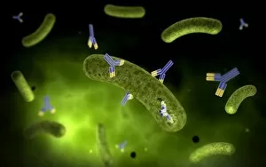
Background imagePathology Collection: Conceptual image of antibody attaching and killing bacteria

Conceptual image of antibody attaching and killing bacteria

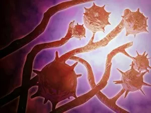
Background imagePathology Collection: Microscopic view of histoplasmosis

Microscopic view of histoplasmosis. Histoplasmosis is an infection in the lungs caused by inhaling the spores of a fungus. This fungus, called Histoplasma capsulatum
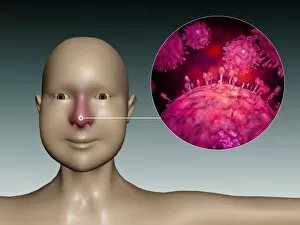
Background imagePathology Collection: Conceptual image of the Human Papilloma Virus

Conceptual image of the Human Papilloma Virus (HPV). HPV is a virus from the papillomavirus family that affects human skin and the moist membranes that line the body

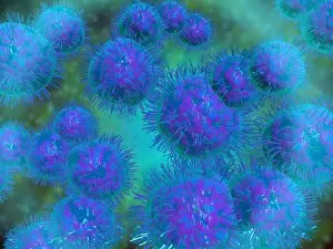
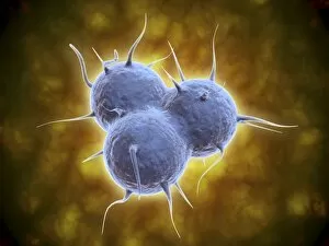
Background imagePathology Collection: Microscopic view of diplococcus bacterium

Microscopic view of diplococcus bacterium

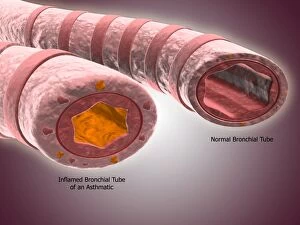
Background imagePathology Collection: Trachea cross-section showing normal and asthmatic bronchiole

Trachea cross-section showing normal and asthmatic bronchiole
Trachea cross-section showing comparison of normal and asthmatic bronchiole

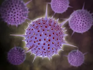
Background imagePathology Collection: Conceptual image of Radiolarians with a skeletal frame

Conceptual image of Radiolarians with a skeletal frame. Radiolarians are tiny protozoans that live in the ocean

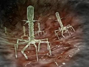
Background imagePathology Collection: Microscopic view of bacteriophages on the surface of a bacteria

Microscopic view of bacteriophages on the surface of a bacteria

Background imagePathology Collection: Common cold influenza virus with microscopic close-up

Common cold influenza virus with microscopic close-up

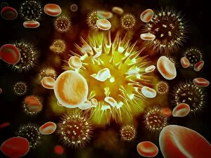
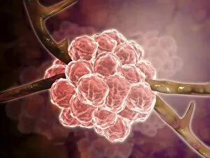
Background imagePathology Collection: Cancer cell with red blood cell flow

Cancer cell with red blood cell flow

Background imagePathology Collection: Electron micrograph of negative-stained Prochlorococcus

Electron micrograph of negative-stained Prochlorococcus. A cyanophage is a virus that infects cyanobacteria

Background imagePathology Collection: Conceptual image of the Hepatitis C virus

Conceptual image of the Hepatitis C virus. Hepatitis C virus is a small, enveloped, positive-sense single-stranded RNA virus of the family Flaviviridae

Background imagePathology Collection: Development of fistula

Development of fistula

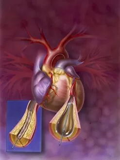
Background imagePathology Collection: Insertion of balloon into atherosclerotic artery

Insertion of balloon into atherosclerotic artery

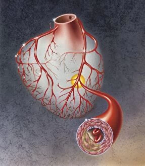
Background imagePathology Collection: Arteries on heart showing atherosclerotic plaque in an artery

Arteries on heart showing atherosclerotic plaque in an artery

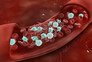
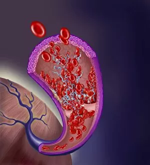
Background imagePathology Collection: Microscopic view of blood clotting inside the artery

Microscopic view of blood clotting inside the artery

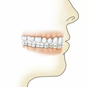
Background imagePathology Collection: Lower gums with braces and plaque on teeth

Lower gums with braces and plaque on teeth

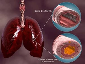
Background imagePathology Collection: Trachea cross-section with lungs showing normal and asthmatic bronchiole

Trachea cross-section with lungs showing normal and asthmatic bronchiole
Trachea cross-section with lungs, showing comparison of normal and asthmatic bronchiole

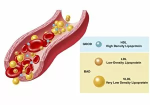
Background imagePathology Collection: Good and bad cholesterol found in blood stream

Good and bad cholesterol found in blood stream

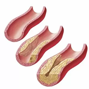
Background imagePathology Collection: Build-up of atherosclerotic plaque in an artery

Build-up of atherosclerotic plaque in an artery

Background imagePathology Collection: Microscopic view of Methicillin-resistant Staphylococcus aureus

Microscopic view of Methicillin-resistant Staphylococcus aureus (MRSA). MRSA is a bacterium responsible for several difficult-to-treat infections in humans

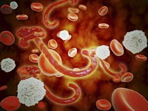
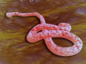
Background imagePathology Collection: Conceptual image of ebola virus in blood stream

Conceptual image of ebola virus in blood stream

Background imagePathology Collection: Microscopic view of Stachybotrys

Microscopic view of Stachybotrys. Stachybotrys is a genus of molds, hyphomycetes or asexually reproducing, filamentous fungi

Background imagePathology Collection: Conceptual image of RNA virus replication

Conceptual image of RNA virus replication

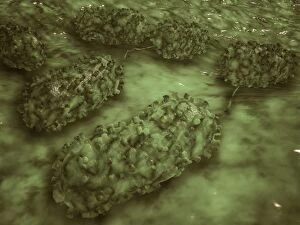
Background imagePathology Collection: Microscopic view of E. coli bacteria cells

Microscopic view of E. coli bacteria cells
Microscopic view of Escherichia coli bacteria cells, commonly known as E. Coli. E. coli is a common type of bacteria that can get into food, such as beef and vegetables

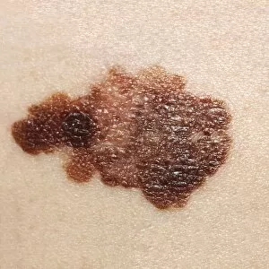
Background imagePathology Collection: Melanoma on a patients skin

Melanoma on a patients skin

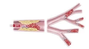
Background imagePathology Collection: Acute coronary syndrome - microvascular obstruction

Acute coronary syndrome - microvascular obstruction

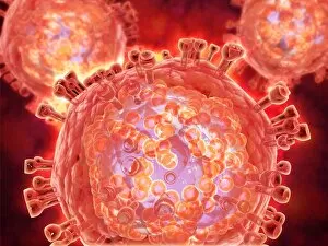
Background imagePathology Collection: Cluster of HIV virus

Cluster of HIV virus. HIV is the human immunodeficiency virus that can lead to acquired immune deficiency syndrom, or AIDS

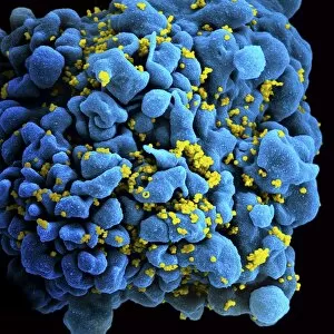
Background imagePathology Collection: Colorized image of HIV-infected H9 T-cell

Colorized image of HIV-infected H9 T-cell

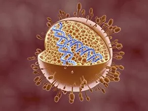
Background imagePathology Collection: Cutaway view of Reoviridae virus showing dna inside

Cutaway view of Reoviridae virus showing dna inside. Reoviruses can affect the gastronintestinal system and respiratory tract

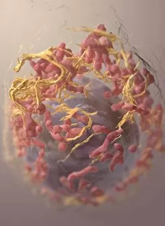
Background imagePathology Collection: 3D structure of melanoma cell

3D structure of melanoma cell
3D structure of a melanoma cell derived by ion abrasion scanning electron microscopy

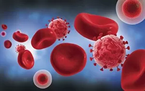
Background imagePathology Collection: Microscopic view of blood cells with virus

Microscopic view of blood cells with virus

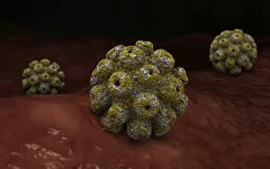
Background imagePathology Collection: Conceptual image of polyomavirus

Conceptual image of polyomavirus

Background imagePathology Collection: Microscopic view of Ebola virus

Microscopic view of Ebola virus (EBOV). EBOV causes extremely severe disease in humans and in non-human primates in the form of viral hemorrhagic fever

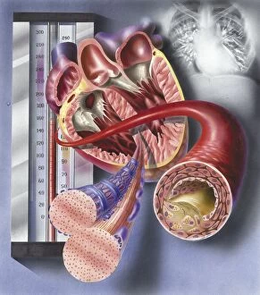
Background imagePathology Collection: Interior view of heart with detail of muscle cells and atherosclerotic artery

Interior view of heart with detail of muscle cells and atherosclerotic artery. Chest x-ray in background

Background imagePathology Collection: Thrombus forming on valve within vein

Thrombus forming on valve within vein


For sale as Licensed Images

Choose your image, Select your licence and Download the media

"Exploring the Intricate World of Pathology: Unveiling the Mysteries Within" Delving into the realm of pathology, we encounter a fascinating array of scientific wonders. From a set of glass eyeballs that have witnessed countless medical marvels to human brain microscope slides revealing intricate neural networks, each specimen holds secrets waiting to be unraveled. Intriguingly, a tobacco hornworm intertwined with its tobacco plant counterpart reminds us of the intricate relationship between organisms and their environment. Similarly, an Alzheimer's brain reveals the devastating effects this disease has on our most precious organ. As we journey through history, we encounter Louis Pasteur, the renowned French microbiologist whose groundbreaking discoveries paved the way for modern medicine. His contributions continue to inspire generations in their pursuit of understanding diseases at their core. Peering through microscopes, we witness captivating views such as that of a microscopic respiratory syncytial virus invading its host or Dohle bodies within blood cells – tiny anomalies holding vital diagnostic clues. The microscopic view of chlamydia serves as a reminder that even seemingly inconspicuous pathogens can wreak havoc on our health. Examining acute promyelocytic leukemia under magnification brings awareness to the battle fought by patients and healthcare professionals alike against this aggressive form of cancer. Meanwhile, glimpsing at sleeping sickness parasites highlights how these minuscule invaders disrupt lives in regions plagued by poverty and limited resources. The sight of liver cirrhosis evokes contemplation about lifestyle choices and their consequences on one's vital organs. In contrast, light micrographs showcasing liver tissue cirrhosis provide insight into cellular changes occurring within this complex disease process. Pathology is not merely confined to textbooks; it is an ever-evolving field where scientists tirelessly strive to decipher nature's enigmatic puzzles. Each slide represents more than just pixels; they hold stories waiting to be told – tales that shed light on diseases afflicting humanity and offer hope for better treatments.