
Pedipalp Collection
Exploring the intricate world of spider anatomy through captivating artwork, we delve into the fascinating realm of pedipalps


For sale as Licensed Images
Choose your image, Select your licence and Download the media
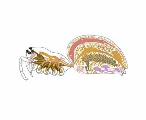
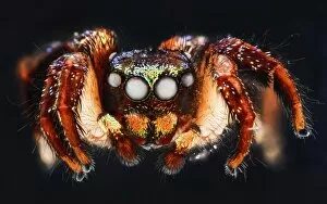
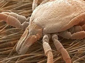
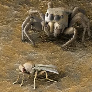
Exploring the intricate world of spider anatomy through captivating artwork, we delve into the fascinating realm of pedipalps. These specialized appendages found in spiders play a crucial role in their survival and hunting techniques. Witness the astonishing details captured in images like Jumping spider C018 / 4312 and Jumping spider C018 / 4470, where these agile creatures showcase their remarkable agility. SEM Z430 / 0436 unveils the mesmerizing complexity of spider mouthparts, highlighting how they are uniquely adapted for feeding and capturing prey. From House spiders to Deer ticks and Sheep ticks, SEM imagery reveals the diversity within this arachnid group. Marvel at Tarantula C016 / 7767 as it displays its formidable pedipalps that aid in grasping and manipulating objects with precision. Meanwhile, Lynx spider C018 / 2420 showcases its elegant pedipalps used for courtship rituals. Intriguingly captured by SEM technology, witness a Spider stalking its unsuspecting prey - an awe-inspiring display of nature's cunning strategies. The detailed close-ups provided by SEM imaging allow us to appreciate every intricate feature that makes spiders such extraordinary creatures. Join us on this visual journey as we uncover the hidden wonders of these eight-legged marvels through stunning SEM images that offer a unique perspective on their anatomy.