Pelvic Collection (#4)

"The Pelvic: A Fascinating Intersection of Art, Anatomy, and Health" The pelvic region has captivated artists and scientists alike throughout history

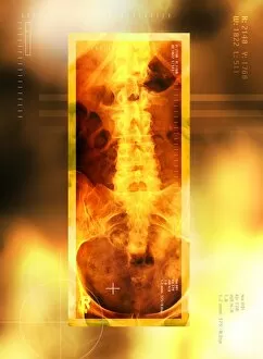
Background imagePelvic Collection: Healthy lower back, X-ray

Healthy lower back, X-ray
Healthy lower back, coloured X-ray. The spine runs down centre. It is made up of vertebrae, block- shaped bones that support and protect the spinal cord (not seen)

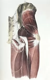
Background imagePelvic Collection: Pelvic spinal nerves

Pelvic spinal nerves. Historical anatomical artwork of the spinal nerves (white) in the human pelvic region. This side view (front of the body at right)

Background imagePelvic Collection: Spinal column

Spinal column. Historical artwork of the bones of the human spine and pelvis, seen from the front. The backbone (vertical)

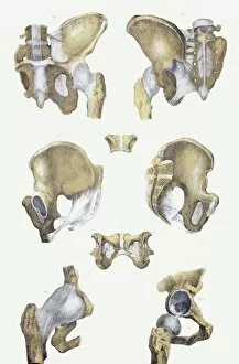
Background imagePelvic Collection: Pelvis bones and ligaments

Pelvis bones and ligaments. Historical anatomical artwork of pelvis bones (yellow) and ligaments (white). Ligaments are bands of fibrous tissue that hold bones together at their joints

Background imagePelvic Collection: Osteoarthritis of hip joints, X-ray

Osteoarthritis of hip joints, X-ray
Hip joint osteoarthritis. Coloured X-ray of the pelvis of a 72-year-old woman showing areas of osteoarthritis of the hip joints (orange)

Background imagePelvic Collection: Osteoporosis

Osteoporosis: illustration of a weakened femoral head (hip joint) at the point of breaking, and the stooped posture of the elderly woman at left

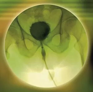
Background imagePelvic Collection: Normal bladder, cystogram

Normal bladder, cystogram
Normal bladder, coloured cystogram (bladder X-ray). The tube running down from the bladder (black) is the catheter used to introduce the contrast medium during retrograde cystography

Background imagePelvic Collection: Fractured hip bone, artwork

Fractured hip bone, artwork
Fractured hip bone. Computer artwork of the bones of the hip showing a break (yellow) in the femoral neck

Background imagePelvic Collection: Pelvic fracture, X-ray

Pelvic fracture, X-ray

Background imagePelvic Collection: Pelvis in Pagets disease, X-ray

Pelvis in Pagets disease, X-ray
Coloured X-ray (front view) of the pelvis in a 60 year old patient showing the mottled and wispy appearance of the bones due to their increased porosity in Pagets disease

Background imagePelvic Collection: Healthy pelvis, 3D CT scan

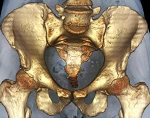
Healthy pelvis, 3D CT scan
Coloured three-dimensional (3D) computed tomography (CT) scan in front view of the healthy pelvis in a 52 year old patient showing the sacrum of the spine, pelvic bones

Background imagePelvic Collection: Pelvis with a leaning posture, X-ray

Pelvis with a leaning posture, X-ray
Coloured X-ray (front view) of the pelvis in a patient with severe knee pain whose walk was affected on one side (at left) and whose posture was lateralised into a sideways leaning gait

Background imagePelvic Collection: Prostate disorder, X-ray urogram

Prostate disorder, X-ray urogram
Coloured X-ray urogram of the pelvis in a 63 year old male patient showing the bladder (light) with the enlarged prostate gland beneath

Background imagePelvic Collection: In-orbit medical report

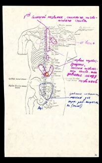
In-orbit medical report. Medical report used in the Soviet space programme: first in 1976 by cosmonaut Vyacheslav Zudov on the Soyuz 23 mission

Background imagePelvic Collection: Bone density scanner, composite image

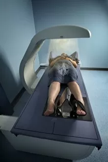
Bone density scanner, composite image
MODEL RELEASED. Bone density scanner. Composite image of a dual energy X-ray absorptiometry (DXA) scanner being used to measure a patients bone mineral density (BMD)

Background imagePelvic Collection: Female pelvis, artwork

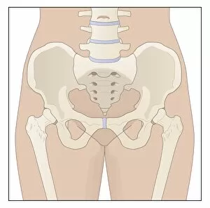
Female pelvis, artwork. The lowest lumbar vertebra of the spinal column (top centre) forms a joint with the sacrum (triangular)

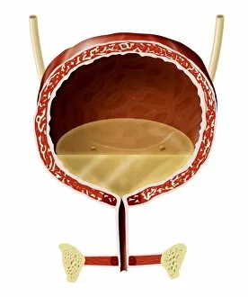
Background imagePelvic Collection: Bladder anatomy, artwork

Bladder anatomy, artwork

Background imagePelvic Collection: Neanderthal skeleton, Kebara Cave, Israel

Neanderthal skeleton, Kebara Cave, Israel
Neanderthal skeleton. Close-up of the skeleton of a Neanderthal (Homo neanderthalensis) found in the Kebara Cave, Israel, in 1982

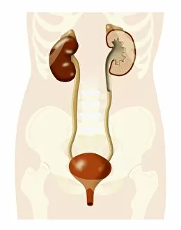
Background imagePelvic Collection: Urinary system, artwork

Urinary system, artwork
Urinary system. Artwork showing the human urinary system on an outline of the human body, showing the location of the kidneys (upper left and upper right) and bladder (lower centre)

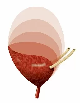
Background imagePelvic Collection: Bladder expansion, artwork

Bladder expansion, artwork
Bladder expansion. Sequential artwork showing the external structure of an expanding bladder. Successive shades of lighter pink show the bladder expanding greatly in volume from its resting state

Background imagePelvic Collection: Main in pain, artwork

Main in pain, artwork
Main in pain, computer artwork

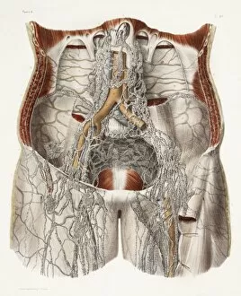
Background imagePelvic Collection: Circulatory system, 19th Century artwork

Circulatory system, 19th Century artwork. Historical hand coloured lithographic print showing the main arteries (red), veins (blue) and lymph vessels (white threads)

Background imagePelvic Collection: Broken hip, X-ray

Broken hip, X-ray

Background imagePelvic Collection: Incontinence implant, X-ray

Incontinence implant, X-ray
Incontinence implant. Coloured frontal X-ray of the pelvic region of a 61-year-old woman with a sacral nerve stimulator (SNS) implanted in the abdomen (dark lines, centre left and right)

Background imagePelvic Collection: Abdominal aortic aneurysm, artwork

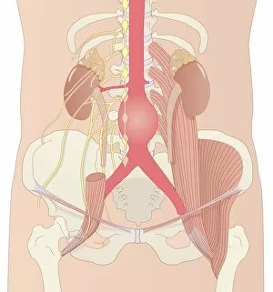
Abdominal aortic aneurysm, artwork
Abdominal aortic aneurysm. Computer artwork showing a large aneurysm (round, centre) in the abdominal section of the aorta

Background imagePelvic Collection: Fractured pelvis, X-ray

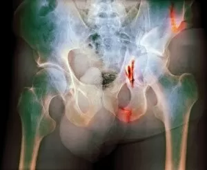
Fractured pelvis, X-ray
Fractured pelvis. Coloured X-ray of fractures (red) in the pelvis. The head of the femur (thigh bone) articulates with the acetabulum to form the hip joint

Background imagePelvic Collection: Ectopic IUD contraceptive, CT scan

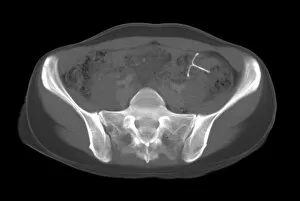
Ectopic IUD contraceptive, CT scan
Ectopic IUD contraceptive. Computed tomography (CT) scan of an axial section through the lower abdomen of a woman, showing an ectopic (abnormally positioned) intrauterine device (IUD, white)

Background imagePelvic Collection: Ectopic IUD contraceptive, X-ray

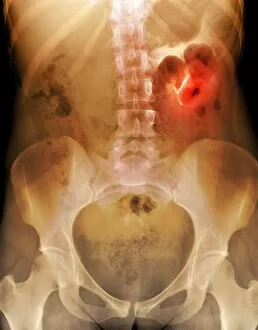
Ectopic IUD contraceptive, X-ray
Ectopic IUD contraceptive. Coloured X-ray of the lower abdomen of a woman, showing an ectopic (abnormally positioned) intrauterine device (IUD, white, highlighted in red)

Background imagePelvic Collection: Dislocated hip replacement, X-ray

Dislocated hip replacement, X-ray
Dislocated hip replacement. Frontal X-ray of the pelvis of a patient with bi-lateral (both sides) total hip replacements. The right joint replacement (right) has become dislocated

Background imagePelvic Collection: Ectopic IUD contraceptive, 3D MRI scan

Ectopic IUD contraceptive, 3D MRI scan
Ectopic IUD contraceptive. Coloured 3D magnetic resonance imaging (MRI) scan of an axial section through the lower abdomen of a woman

Background imagePelvic Collection: Skeleton and ligaments, artwork

Skeleton and ligaments, artwork
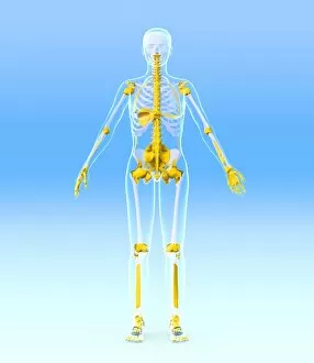
Female skeleton, computer artwork. The human skeleton has 206 bones. The skull (at top) protects the brain. The ribs of the chest (at upper centre) enclose the heart and lungs

Background imagePelvic Collection: Abdominal organs, anatomical artwork

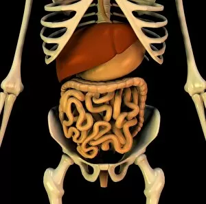
Abdominal organs, anatomical artwork. The organs are shown within the surrounding bones, including the pelvis (bottom) and the rib-cage (top)

Background imagePelvic Collection: Neck of femur fracture, X-ray

Neck of femur fracture, X-ray
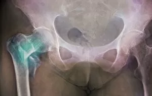
Coloured X-ray (front view) of the pelvis of a patient, showing a hip fracture (at left) to the neck of the thigh bone (femur)

Background imagePelvic Collection: Dislocated hip prosthesis, X-ray

Dislocated hip prosthesis, X-ray
Coloured X-ray (front view) of the pelvis of a patient, showing the dislocation of an artificial (prosthetic) total hip joint replacement


For sale as Licensed Images

Choose your image, Select your licence and Download the media

"The Pelvic: A Fascinating Intersection of Art, Anatomy, and Health" The pelvic region has captivated artists and scientists alike throughout history. From ancient historical artwork depicting the cardiovascular system's connection to the pelvis to modern depictions of hip replacement surgeries, this area holds immense significance. One common ailment that affects the pelvic region is osteoarthritis of the hip. X-ray images reveal its impact on the bones, prompting advancements in treatments like hip replacements. These procedures have revolutionized healthcare by restoring mobility and alleviating pain for countless individuals. Another condition associated with the pelvis is a slipped disc. Artists have beautifully portrayed this issue through intricate artworks showcasing slipped intervertebral discs. Such visuals help medical professionals understand and treat this painful condition effectively. Delving deeper into anatomy, X-rays provide detailed views of hip bones from various angles. The complexity of these structures becomes evident as we explore their connections to muscles such as Levator ani - an engraving displaying a side view of the pelvis highlights this relationship. The wonderment surrounding life begins within the womb, where diagrams depict foetuses developing within their mothers' pelvises. This glimpse into human reproduction showcases how essential a healthy pelvic structure is for nurturing new life. Artists have also contributed to our understanding of skeletal anatomy by creating stunning artwork portraying human skeletons in intricate detail. By studying these pieces, we gain insights into how different parts interact within the body's framework. Amidst all these scientific explorations lies an unexpected surprise – a blacktip reef shark. Though not directly related to human anatomy or health concerns involving the pelvic region, it serves as a reminder that nature's wonders extend beyond our own bodies. Returning back to humans, 1831 artwork focuses on another crucial aspect: pelvic-femoral muscles. Understanding their functions aids in comprehending movement patterns and potential issues that may arise when they are compromised. Lastly, artistic representations shed light on the complexity of hip joint bones and their anatomy.