
Phagocyte Collection
"Phagocytes: The Mighty Protectors of our Immune System" In the intricate world of immunology, dendritic cells take center stage as the orchestrators of immune responses


For sale as Licensed Images
Choose your image, Select your licence and Download the media
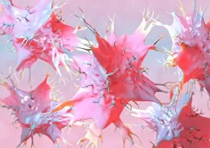
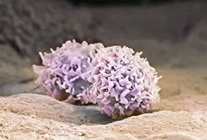
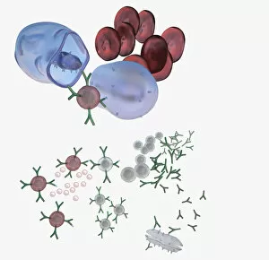
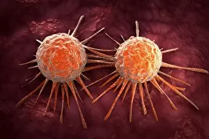
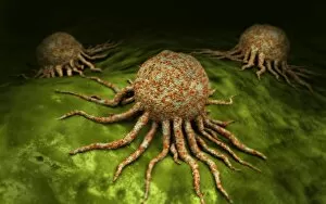
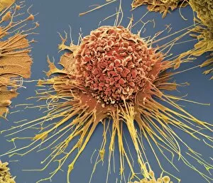
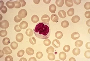
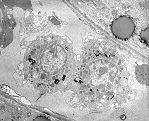
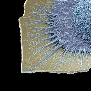
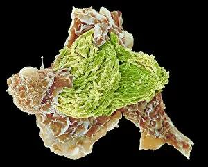
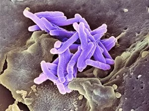
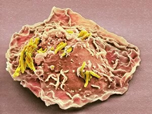
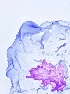
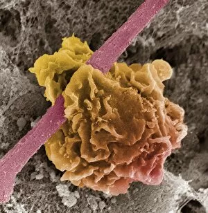
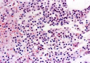
"Phagocytes: The Mighty Protectors of our Immune System" In the intricate world of immunology, dendritic cells take center stage as the orchestrators of immune responses. Their fascinating structure and function are beautifully depicted in artwork that captures their complexity. Activated macrophages, captured through scanning electron microscopy (SEM C015 / 6375), showcase their formidable power to engulf and destroy invading pathogens. These warriors play a crucial role in defending against diseases like multiple sclerosis, as seen in another SEM image. Dendritic cells once again grace us with their presence in stunning artwork, reminding us of their pivotal role in presenting antigens to activate other immune cells. Meanwhile, transmission electron microscopy (TEM) reveals the intricacies of macrophage cells at a microscopic level. An illustration depicting an immune response triggered by a microbe showcases the chain reaction involving defensive white blood cells. This captivating image highlights how phagocytes work together harmoniously to protect our bodies from harm. Conceptual images portraying cancer viruses serve as a stark reminder that phagocytes also combat malignant threats within our own bodies. These powerful defenders relentlessly seek out and eliminate cancerous invaders on a microscopic scale. Microscopic views provide glimpses into the world of phagocytic macrophages working tirelessly to rid our bodies of foreign substances. Whether it's observing individual macrophages or witnessing them gather as a group, these images demonstrate their unwavering dedication to maintaining our health. A striking 3D rendering visualizes macrophage phagocytosis - capturing the moment when these remarkable cells engulf and digest harmful particles with precision and efficiency. In this captivating journey through imagery, we witness the extraordinary capabilities of phagocytes – nature's ultimate guardians standing tall against infections and disease while preserving our well-being.