Phalanx Collection (#10)

"Unveiling the Intricacies of the Phalanx: From Normal Foot to Inner Ankle Ligaments" In this captivating journey through human anatomy

Background imagePhalanx Collection: Dislocated toe, X-ray

Dislocated toe, X-ray
Dislocated toe. Coloured X-ray of the foot of a 49 year old woman with a dislocated toe. The dislocated joint is shown in yellow. It is the fourth proximal interphalangeal joint

Background imagePhalanx Collection: Thalassaemia, X-ray

Thalassaemia, X-ray
Thalassaemia. Coloured hand X-ray of a patient with thalassaemia, also known as Cooleys anaemia. The bones of the hand show demineralization (lighter patches)

Background imagePhalanx Collection: Hand bones

Hand bones. Computer artwork of a healthy hand and wrist joint. There are eight wrist (carpal) bones (bottom right). The carpal bones articulate with the bones of the forearm

Background imagePhalanx Collection: Foot bones

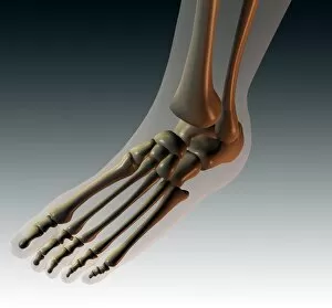
Foot bones, computer artwork. The toes are made up of the phalanx bones (phalanges), two for the big toe (lower left) and three for the others

Background imagePhalanx Collection: Hand bones and ligaments

Hand bones and ligaments. Historical anatomical artwork of the bones (yellow) & ligaments (white) of the hand. Ligaments are bands of fibrous tissue that hold bones together at their joints

Background imagePhalanx Collection: Pinned finger fracture, X-ray

Pinned finger fracture, X-ray
Pinned broken finger. Coloured X-ray of a pinned fracture (red area) of the index finger. Five metal pins (dark brown) are holding the bone (the first phalanx)

Background imagePhalanx Collection: Dislocated finger

Dislocated finger

Background imagePhalanx Collection: Finger inflammation, X-ray

Finger inflammation, X-ray
Inflamed finger. Coloured X-ray of a pointing finger, with an inflammatory sore (felon, lower right). The middle finger bone (phalanx, centre) has also been infected and become thinned

Background imagePhalanx Collection: Arthritic hand

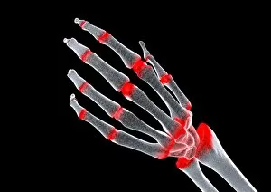
Arthritic hand, computer artwork. The finger and wrist joints are inflamed and swollen. Rheumatoid arthritis is caused by the immune system attacking the bodys own tissues

Background imagePhalanx Collection: Arthritic hand, conceptual artwork

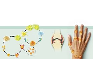
Arthritic hand, conceptual artwork. Arthritis is a degenerative disease that results in the loss of cartilage between joints (red), causing pain and inflammation

Background imagePhalanx Collection: Rheumatoid arthritis mechanism, artwork

Rheumatoid arthritis mechanism, artwork
Rheumatoid arthritis mechanism. Artwork showing various aspects of rheumatoid arthritis, the inflammation of the joints due to attack by the bodys own immune system

Background imagePhalanx Collection: The bones of the foot

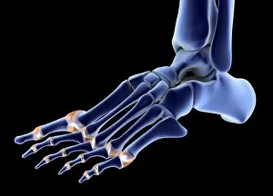
The bones of the foot
3D medical illustration of a left side view of the bones of the left foot, showing the metatarsal phalangeal joints

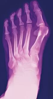
Background imagePhalanx Collection: Bunion, X-ray

Bunion, X-ray
Bunion. Coloured X-ray of a bunion (hallux valgus, upper right), the swelling of the joint between the big toe and the first metatarsal bone in the foot

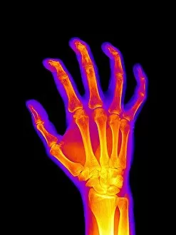
Background imagePhalanx Collection: Arthritic hand, X-ray

Arthritic hand, X-ray
Arthritic hand, coloured X-ray

Background imagePhalanx Collection: Fingertip laceration injuries, X-rays

Fingertip laceration injuries, X-rays
Fingertip laceration injuries, coloured X-rays. Both X-rays show the same hand, which suffered severe lacerations (blunt trauma cuts) to the tips of the middle fingers (green)

Background imagePhalanx Collection: Amputated fingertip, X-ray

Amputated fingertip, X-ray
Amputated fingertip. Coloured X-ray of the 4th and 5th fingers of a hand where the end of the 5th (little) finger has been amputated (red)

Background imagePhalanx Collection: Hand anatomy, artwork

Hand anatomy, artwork
hand, bone, skeleton

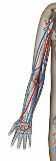
Background imagePhalanx Collection: Arm blood vessels, artwork

Arm blood vessels, artwork. Within the outlined body shape (grey) are the bones (also grey) and the arteries (red) and veins (blue). In the upper arm, the bone is the humerus

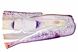
Background imagePhalanx Collection: Human finger, longitudinal section

Human finger, longitudinal section
Human finger. Light micrograph of a longitudinal section through a finger of a human infant. This shows the bones inside the finger (here, the 1st and 2nd phalanges)

Background imagePhalanx Collection: Human skeleton, artwork

Human skeleton, artwork
Human skeleton. Computer artwork of a human skeleton, with a close-up of its hand. The skull (upper centre) and rib cage (centre and lower centre) can also be seen


For sale as Licensed Images

Choose your image, Select your licence and Download the media

"Unveiling the Intricacies of the Phalanx: From Normal Foot to Inner Ankle Ligaments" In this captivating journey through human anatomy, we explore the fascinating world of the phalanx. Starting with a normal foot X-ray, we delve into the intricate layers that make up this vital part of our body. The human foot anatomy reveals a mesmerizing tapestry of skin, veins, arteries, muscles, and bones working in harmony. Moving on to outer ankle ligaments depicted in artwork C013/4452, we witness their strength and resilience as they support our every step. Similarly, inner ankle ligaments portrayed in artwork C013/4451 showcase their crucial role in maintaining stability and balance. Venturing beyond mere physicality, palmistry introduces us to a planetary and zodiacal diagram that adds an element of mystique to our exploration. Meanwhile, Francisco Franco's "Diario de una Bandera" cover reminds us of historical significance tied to symbols like flags. As we dive deeper into history's annals, iconic battles come alive before our eyes. From "The Battle of Waterloo, " immortalized on canvas by skilled artists capturing its intensity; to Pausanias' grandeur illustrated in Hutchinsons History; or even comparing hand bones across diverse mammals - each depiction unveils unique aspects related to warfare or evolution. Finally reaching ancient times when military formations reigned supreme comes "The Phalanx attacking the Centre on the Hydaspes. " This lithograph transports us back to those fierce encounters where unity and coordination were paramount for success. And who can forget "The Battle of Marathon, " another pivotal moment etched forever within Hutchinsons History? Concluding with a colored engraving showcasing "The Macedonians, " we realize how these warriors left an indelible mark on history through their strategic use of phalanxes.