Physiological Collection (#15)

"Exploring the Intricacies of Physiology: From Airpumps to Blood Cells" Delving into the world of physiology

Background imagePhysiological Collection: Melatonin brain function, artwork

Melatonin brain function, artwork
Melatonin brain function. Artwork showing how the hormone melatonin (blue spheres) is secreted in the brain and helps to control the bodys biological clock

Background imagePhysiological Collection: Heart valves, artwork

Heart valves, artwork
Heart valves. Cutaway artwork of the inside of the heart, showing the various valves. The first two artworks (left and centre) show the atrioventricular valves between the hearts atria (top)

Background imagePhysiological Collection: Nerve cell and axon, diagram

Nerve cell and axon, diagram
Nerve cell and axon. Diagram showing how the input to a nerve cell (neuron) through dendrites (shown by arrows at upper left) is transmitted (yellow arrow) along the nerve cells axon (across bottom)

Background imagePhysiological Collection: Intestinal anatomy, artwork

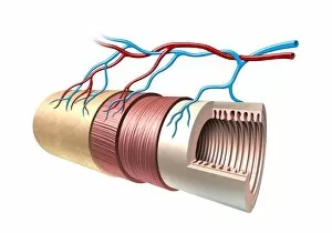
Intestinal anatomy, artwork
Intestinal anatomy. Computer artwork showing the layers of the small intestine. The central space (lumen) is surrounded by the mucosa (beige, folded), which has numerous folds (villi)

Background imagePhysiological Collection: Respiratory tract, artwork

Respiratory tract, artwork
Respiratory tract. Computer artwork showing the various stages and structures of the human respiratory tract. Air is drawn in through the oral and/or nasal cavities, and passes down the trachea

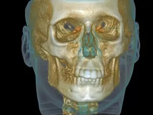
Background imagePhysiological Collection: Normal head, cone beam CT scan

Normal head, cone beam CT scan. Front view of a dental patients head. The scan was produced using a cone beam x-ray computed tomography (CT) scanner

Background imagePhysiological Collection: Abdominal aortic aneurysm, artwork

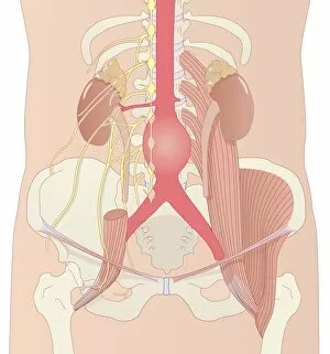
Abdominal aortic aneurysm, artwork
Abdominal aortic aneurysm. Computer artwork showing a large aneurysm (round, centre) in the abdominal section of the aorta

Background imagePhysiological Collection: Shin splints, artwork

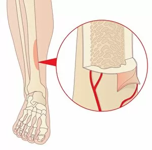
Shin splints, artwork
Shin splints. Computer artwork of a lower leg showing an area of the tibia (shin bone) affected by medial tibial stress syndrome, or shin splints

Background imagePhysiological Collection: Stem cell therapy, artwork

Stem cell therapy, artwork
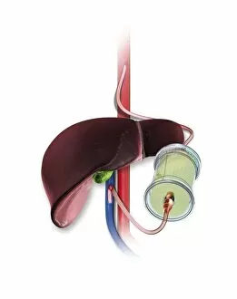
Stem cell therapy. Artwork showing stem cells (in green cylinder) being added to a patients bloodstream in the region of the liver (brown)

Background imagePhysiological Collection: Penguins mating, artwork

Penguins mating, artwork. The penguins reproductive organs are shown here, with the male on top and the female on the bottom

Background imagePhysiological Collection: Edoardo Perroncito, Italian physician

Edoardo Perroncito, Italian physician
Edoardo Perroncito (1847Ôé¼" 1936), Italian physician, veterinarian and parasitologist. Perroncito is credited, along with Luigi Pagliani (1847Ôé¼" 1931)

Background imagePhysiological Collection: Retinal rod cell anatomy, diagram

Retinal rod cell anatomy, diagram

Background imagePhysiological Collection: Bowmans capsule, diagram

Bowmans capsule, diagram
Bowmans capsule. Diagram showing the anatomical structure of a Bowmans capsule and glomerulus in a kidney

Background imagePhysiological Collection: Lung alveolus, artwork

Lung alveolus, artwork
Lung alveolus, computer artwork. Alveoli are the site of gas exchange in the lungs of mammals, where oxygen is taken up and carbon dioxide released

Background imagePhysiological Collection: Vestibular ciliated cells, diagram

Vestibular ciliated cells, diagram
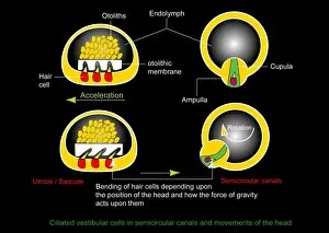
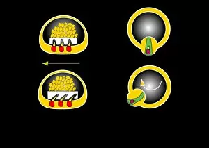
Vestibular ciliated cells. Diagram of the anatomical structure and function of the vestibular ciliated cells found in the inner ear that help sense and control of balance

Background imagePhysiological Collection: Human lungs

Human lungs. Anatomical artwork of the human lungs and respiratory system. The heart (which has been removed here) is located between the lungs

Background imagePhysiological Collection: Brain anatomy, diagram

Brain anatomy, diagram
Brain anatomy. Diagram of the underside (ventral aspect) of the brain, showing the anatomical structure of the various components

Background imagePhysiological Collection: Milk-producing cell, artwork

Milk-producing cell, artwork
Milk-producing cell. Artwork of the anatomical structure of a lacteal (milk-producing) cell. Normal cell organelles include the nucleus (black, centre), the golgi apparatus (white)

Background imagePhysiological Collection: Taste bud anatomy, diagram

Taste bud anatomy, diagram
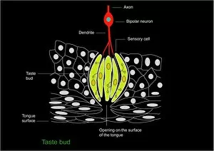
Taste bud anatomy. Diagram of the anatomical structure of a taste bud on a tongue. The surface of the tongue is at bottom, with the tastebud (rounded structure) at centre

Background imagePhysiological Collection: Semicircular canal, artwork

Semicircular canal, artwork
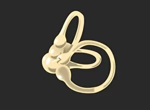
Semicircular canal. Artwork of the anatomical structure of the semicircular duct or canal, a structure in the inner ear that help detect head movements

Background imagePhysiological Collection: Semicircular canal, diagram

Semicircular canal, diagram
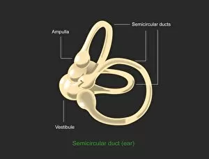
Semicircular canal. Diagram of the anatomical structure of the semicircular duct or canal, a structure in the inner ear that help detect head movements

Background imagePhysiological Collection: Vestibular ciliated cells, artwork

Vestibular ciliated cells, artwork
Vestibular ciliated cells. Artwork of the anatomical structure and function of the vestibular ciliated cells found in the inner ear that help sense and control of balance

Background imagePhysiological Collection: Elie Metchnikoff, Russian biologist

Elie Metchnikoff, Russian biologist
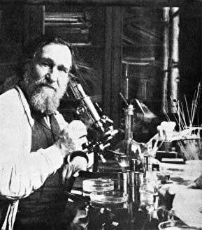
Elie Metchnikoff (1845-1916), Russian biologist and Nobel laureate. Metchnikoff, also known as Ilya Ilyich Mechnikov, is best known for his pioneering research into the immune system

Background imagePhysiological Collection: Capillary, artwork

Capillary, artwork
Capillary. Artwork showing the anatomical structure of a capillary seen in cross-section

Background imagePhysiological Collection: Retinal rod cell anatomy, artwork

Retinal rod cell anatomy, artwork
Retinal rod cell anatomy. Artwork of the external (left) and internal (right) anatomical structure of a retinal cell. These cells are found in the retina that lines the back of the eyes

Background imagePhysiological Collection: Muscle contraction, artwork

Muscle contraction, artwork
Muscle contraction. Artwork showing the mechanism for muscle contraction in skeletal and cardiac muscle. At top right troponin (T, C, I) is bound to tropomyosin (red)

Background imagePhysiological Collection: Nerve cell with electrical sparks

Nerve cell with electrical sparks
Nerve cell. Conceptual computer artwork of a nerve cell, also called a neuron emitting electrical sparks. Neurons are responsible for passing information around the central nervous system (CNS)

Background imagePhysiological Collection: Milk-producing cell, diagram

Milk-producing cell, diagram
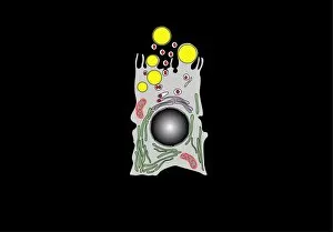
Milk-producing cell. Diagram of the anatomical structure of a lacteal (milk-producing) cell. Normal cell organelles include the nucleus (black, centre), the golgi apparatus (white)

Background imagePhysiological Collection: Glomerular anatomy, diagram

Glomerular anatomy, diagram
Glomerular anatomy. Diagram of the anatomical structure of a glomerulus in a kidney

Background imagePhysiological Collection: Bronchus and bronchioles, artwork

Bronchus and bronchioles, artwork
Bronchus and bronchioles. Artwork of the anatomical structure of the bronchi and their bronchioles, the main aspects of the respiratory system

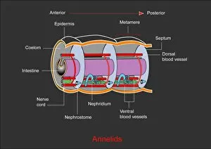
Background imagePhysiological Collection: Annelid worm anatomy

Annelid worm anatomy. Computer artwork showing the internal anatomy of 3 segments (metameres) from an annelid (segmented) worms body

Background imagePhysiological Collection: Activated platelets, artwork

Activated platelets, artwork
Activated platelets. Artwork of platelets (thrombocytes) that have been activated. Platelets are part of the blood. When a blood vessel is damaged

Background imagePhysiological Collection: Human fertilisation, artwork

Human fertilisation, artwork
Human fertilization. Artwork of a single sperm cell (spermatozoon) attempting to fertilise a human egg cell (ovum, round, lower right). The sperm cell consists of a head section and a tail section

Background imagePhysiological Collection: Spallanzanis digestion experiment

Spallanzanis digestion experiment. Historical artwork of an experiment on digestion carried out by the Italian biologist Lazzaro Spallanzani (1729-1799)

Background imagePhysiological Collection: Neuromuscular junctions, artwork

Neuromuscular junctions, artwork
Neuromuscular junctions. Computer artwork of junctions between nerves (thread-like objects) and a muscle (purple surface)

Background imagePhysiological Collection: Football kick, skeleton artwork

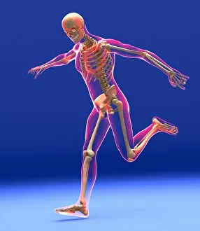
Football kick, skeleton artwork
Football kick. Computer artwork showing a skeleton in the process of kicking a football (not seen). The outline of the body can also be seen

Background imagePhysiological Collection: Synapses, artwork

Synapses, artwork
Synapses. Computer artwork of synapses, the junctions between the ends (green, swollen) of two nerve cells (neurons). Signals are passed along nerve cells in the form of an electrical impulse

Background imagePhysiological Collection: African elephant eye and skin

African elephant eye and skin
Elephant eye and skin. Close up of the eye of an African elephant (Loxodonta africana). The long eyelashes are to protect the eyes from dust and insects

Background imagePhysiological Collection: Albrecht von Haller, Swiss anatomist

Albrecht von Haller, Swiss anatomist
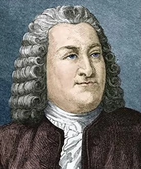
Albrecht von Haller (1708-1777), Swiss anatomist and physiologist. Von Haller worked on the nervous system, and recognised the tendency of muscle fibres to contract when stimulated

Background imagePhysiological Collection: Lung alveoli and red blood cells, TEM

Lung alveoli and red blood cells, TEM
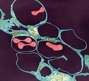
Lung alveoli and red blood cells. Coloured transmission electron micrograph (TEM) of a section through lung alveoli (blue) and red blood cells (red)

Background imagePhysiological Collection: Lung alveoli and blood cells, TEM

Lung alveoli and blood cells, TEM
Lung alveoli and blood cells. Coloured transmission electron micrograph (TEM) of a section through lung alveoli and blood cells

Background imagePhysiological Collection: Asthma and mucus overproduction, artwork

Asthma and mucus overproduction, artwork
Asthma and mucus overproduction. Artwork showing how the bodys immune response causes the inflammation seen in asthma. The chest of a human figure is at top right

Background imagePhysiological Collection: Kidney glomerulus surface, SEM

Kidney glomerulus surface, SEM
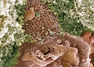
Kidney glomerulus surface. Coloured scanning electron micrograph (SEM) of podocyte cells (orange) on the surface of a kidney glomerulus. These have branching processes (pedicles) that interlock

Background imagePhysiological Collection: William Harvey lecturing on circulation

William Harvey lecturing on circulation
William Harvey (1578-1657), English physician, dissecting an animal during a lecture on the circulation of blood. By 1616 Harvey had dissected 80 species of animals

Background imagePhysiological Collection: William Harvey, English physician

William Harvey, English physician
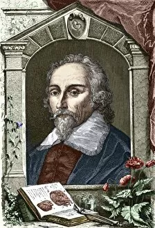
William Harvey (1578-1657), English physician. Harvey is considered the founder of modern physiology for his work on the circulation of the blood

Background imagePhysiological Collection: Organic food, conceptual image

Organic food, conceptual image. Cut-out diagram of a mans head and digestive system, attached to a wooden background. Fruit and vegetables, and plastic sheep and pigs

Background imagePhysiological Collection: Insulin-like growth 1 factor molecule

Insulin-like growth 1 factor molecule
Insulin-like growth factor 1 molecule. Computer model showing the structure of a molecule of the hormone insulin-like growth factor 1 (IGF-1)

Background imagePhysiological Collection: Thyroid-stimulating hormone molecule

Thyroid-stimulating hormone molecule. Computer model showing the structure of a molecule of thyroid stimulating hormone (TSH)


For sale as Licensed Images

Choose your image, Select your licence and Download the media

"Exploring the Intricacies of Physiology: From Airpumps to Blood Cells" Delving into the world of physiology, we uncover a fascinating array of scientific discoveries and visual wonders. Joseph Wright's masterpiece, "The Airpump, " captures the essence of early physiological experiments, showcasing humanity's thirst for knowledge. Intricate synapse nerve junctions depicted through Transmission Electron Microscopy (TEM) reveal the intricate communication network within our bodies. These microscopic connections enable seamless transmission of signals between neurons, shaping our thoughts and actions. Moving upwards to the head, we encounter an astonishing view of the muscular system. The complexity and precision required for even simple movements become apparent as ligaments intertwine with bones in perfect harmony. Venturing deeper into female physiology, a Scanning Electron Microscope (SEM) reveals the uterus lining during menstruation. This glimpse into nature's cyclical process sheds light on one aspect of womanhood that has both fascinated and perplexed scientists throughout history. Stepping away from biology but still within the realm of physiology, lie detector tests emerge as tools to decipher truth from deception. By monitoring physiological responses such as heart rate and perspiration levels, these tests attempt to unravel hidden truths buried within our bodies. Returning to cellular structures through TEM imaging once again unveils rough endoplasmic reticulum - a vital component responsible for protein synthesis in cells. Its intricate web-like structure showcases nature's elegance at work. Tracing back centuries ago brings us to Descartes' optics theory from the 17th century - an exploration into how vision is perceived by our eyes and interpreted by our brains. This groundbreaking theory laid foundations for understanding human perception today. Transitioning towards physicality itself leads us to skeletons intertwined with ligaments - providing structural support while allowing flexibility in movement. These silent heroes ensure smooth locomotion throughout life's journey.