Planktonic Collection (#5)

"Exploring the Intricate World Life: Unveiling the Hidden Beauty through SEM Imaging" Diatoms, one of the most fascinating organisms in pond life

Background imagePlanktonic Collection: Plankton cell wall, SEM

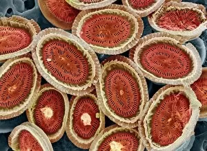
Plankton cell wall, SEM
Plankton cell wall. Coloured scanning electron micrograph (SEM) of the geometrical plates making up the mineralised cell wall of a planktonic alga (Coronosphaera mediterranea)

Background imagePlanktonic Collection: Krill head, SEM

Krill head, SEM
Antarctic krill head (Euphausia superba), coloured scanning electron micrograph (SEM). This animal is a marine crustacean and an important food for many marine animals

Background imagePlanktonic Collection: Rotifer worm, light micrograph

Rotifer worm, light micrograph
Rotifer worm. Light micrograph of the rotifer worm Platyas quadricornis. This free-swimming, aquatic micro-organism has a body which is divided into three sections

Background imagePlanktonic Collection: Cyclops sp. copepod, SEM

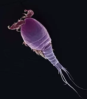
Cyclops sp. copepod, SEM
Cyclops sp. copepod. Coloured scanning electron micrograph (SEM) of a freshwater copepod (Cyclops sp.). The head of the animal is at upper left

Background imagePlanktonic Collection: Rotifer colony

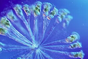
Rotifer colony. Light micrograph of a colony of Conochilus hippocrepis rotifer worms. The colony is composed of between 50 and 100 individual worms, attached at the base of their feet

Background imagePlanktonic Collection: Prorocentrum dinoflagellates, SEM

Prorocentrum dinoflagellates, SEM
Three Prorocentrum dinoflagellates (blue), coloured scanning electron micrograph (SEM). Dinoflagellates are unicellular protozoans

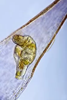
Background imagePlanktonic Collection: Rotifer feeding, light micrograph

Rotifer feeding, light micrograph
Rotifer feeding. Light micrograph of a rotifer (centre) feeding on an alga (green, left). Rotifers (phylum Rotifera) are microscopic aquatic animals that are related to roundworms

Background imagePlanktonic Collection: Copepod with eggs

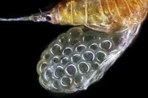
Copepod with eggs. Free-swimming copepod carrying a cluster of eggs (red, left) under its tail. Copepods are microscopic aquatic crustaceans found in waters around the world

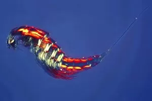
Background imagePlanktonic Collection: Copepod

Copepod. Copepods are microscopic aquatic crustaceans found in waters around the world. They may either be free living or parasitic on other organisms

Background imagePlanktonic Collection: Foraminiferan, light micrograph

Foraminiferan, light micrograph. Foraminifera are marine single-celled protozoa that construct and inhabit shells composed of several chambers

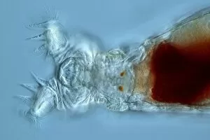
Background imagePlanktonic Collection: Philodina rotifer, light micrograph

Philodina rotifer, light micrograph
Philodina rotifer. Light micrograph of a Philodina roseola rotifers head, showing its mouthparts (left). Rotifers are microscopic aquatic animals that are related to roundworms

Background imagePlanktonic Collection: Diatoms, light micrograph

Diatoms, light micrograph. Diatoms are a group of photosynthetic, single-celled algae containing about 10, 000 species. They form an important part of the plankton at the base of the marine

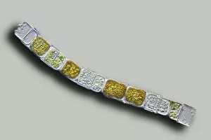
Background imagePlanktonic Collection: Melosira sp diatoms, light micrograph

Melosira sp diatoms, light micrograph
Diatoms. Light micrograph of Melosira monoliformis freshwater diatoms. Diatoms are a group of photosynthetic, single-celled algae containing about 10, 000 species

Background imagePlanktonic Collection: Pleurosigma sp diatoms, light micrograph

Pleurosigma sp diatoms, light micrograph
Diatoms. Differential interference contrast micrograph of Pleurosigma angulatum marine diatoms. Diatoms are a group of photosynthetic, single-celled algae containing about 10, 000 species

Background imagePlanktonic Collection: Fossil diatoms, light micrograph

Fossil diatoms, light micrograph. Diatoms are a group of photosynthetic, single-celled algae containing about 10, 000 species

Background imagePlanktonic Collection: Rotifer foot, light micrograph

Rotifer foot, light micrograph. Rotifers are microscopic aquatic animals that are related to roundworms. The hair-like cilia surrounding the rotifers mouth (right) are used to trap food particles

Background imagePlanktonic Collection: Pleurosigma sp diatom, light micrograph

Pleurosigma sp diatom, light micrograph
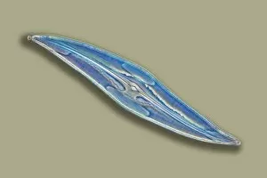
Diatom. Light micrograph of Pleurosigma angulatum marine diatoms. Diatoms are a group of photosynthetic, single-celled algae containing about 10, 000 species

Background imagePlanktonic Collection: Hydromedusa

Hydromedusa. Medusa stage of a Halitholus sp. hydrozoan. Hydromedusae are the sexually reproductive stage of hydrozoan cnidarians

Background imagePlanktonic Collection: Diatom shell, SEM

Diatom shell, SEM
Diatom shell. Coloured scanning electron micrograph (SEM) of the shell of a Biddulphia sp. diatom. The diatoms are a group of photosynthetic, single-celled algae containing about 10, 000 species

Background imagePlanktonic Collection: Dinoflagellate, SEM

Dinoflagellate, SEM
Dinoflagellate. Coloured scanning electron micrograph (SEM) of the dinoflagellate Prorocentrum balticum. Dinoflagellates are unicellular (single-celled) protozoans

Background imagePlanktonic Collection: Marine protozoa shells, SEM

Marine protozoa shells, SEM
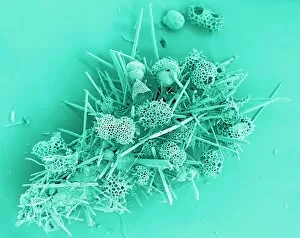
Marine protozoa shells, coloured scanning electron micrograph (SEM). The shells (tests) seen here include those of foraminifera, as well as coral spicules, radiolarian tests and cocccoliths

Background imagePlanktonic Collection: Selection of diatoms, SEM

Selection of diatoms, SEM
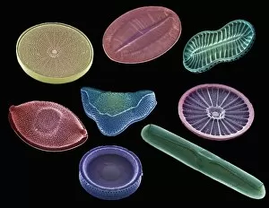
Selection of diatoms, coloured scanning electron micrograph (SEM). Diatoms are single-celled photosynthetic algae, of which there are about 100, 000 species

Background imagePlanktonic Collection: Coscinodiscus diatom, SEM

Coscinodiscus diatom, SEM
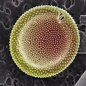
Coscinodiscus diatom. Coloured scanning electron micrograph (SEM) of a Coscinodiscus diatom (red). This is a marine diatom

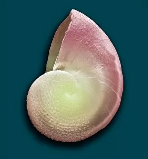
Background imagePlanktonic Collection: Gastropod mollusc larva, SEM

Gastropod mollusc larva, SEM
Gastropod mollusc larva. Coloured scanning electron micrograph (SEM) of the shell of a early stage in the larval development of a gastropod mollusc


For sale as Licensed Images

Choose your image, Select your licence and Download the media

"Exploring the Intricate World Life: Unveiling the Hidden Beauty through SEM Imaging" Diatoms, one of the most fascinating organisms in pond life, reveal their intricate structures under scanning electron microscopy (SEM). The diatom cell wall, composed of silica, showcases a mesmerizing array of patterns and shapes. These microscopic algae play a crucial role in aquatic ecosystems as primary producers. Delving deeper into this captivating realm, we encounter dinoflagellates - another group organisms that exhibit stunning diversity when observed under SEM. Their unique features and complex structures are unveiled through high-resolution imaging techniques. The diatom frustule is an exquisite example of nature's artistry. Its delicate symmetry and intricate designs serve both functional and aesthetic purposes. SEM allows us to appreciate the beauty hidden within these tiny shells. Moving beyond diatoms, we come across foraminifer models - marine protists with elaborate calcium carbonate shells. Examining them using SEM provides insights into their evolutionary adaptations and ecological significance. With each image captured by SEM, we gain a deeper understanding of the complexity and diversity present within planktonic communities. From diatoms to dinoflagellates to foraminifers – these microorganisms shape our planet's ecosystems in ways often unnoticed by human eyes. Through scientific exploration aided by advanced imaging technologies like SEM, we unlock secrets held within these minuscule creatures' world, and is an awe-inspiring journey that reveals not only their structural intricacies but also highlights their vital contributions to Earth's biodiversity and overall environmental health. So next time you gaze upon a tranquil pond or delve into studies on planktonic life, remember the unseen wonders that lie beneath its surface – waiting to be discovered through tools like SEM that bring forth the astonishing beauty found within every diatom shell or algal cell wall.