
Plant Anatomy Collection (#2)
"Exploring the Intricate World of Plant Anatomy: A Fascinating Journey through Micrographs and Artwork" Delve into the hidden wonders with these captivating images


For sale as Licensed Images
Choose your image, Select your licence and Download the media
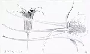
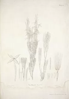
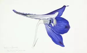
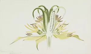
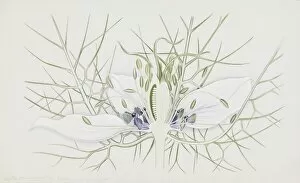
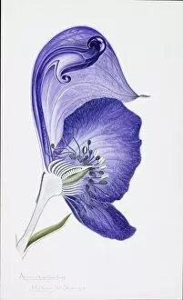
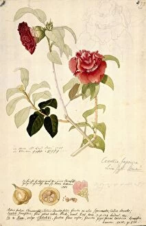
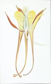
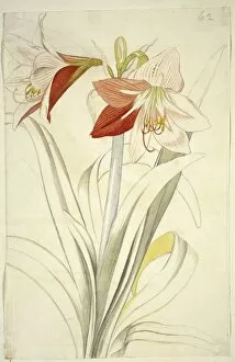
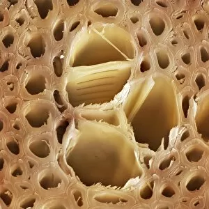
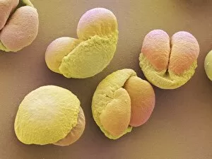
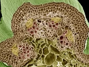
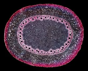
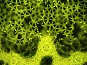
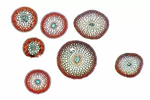
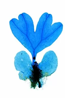
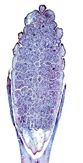
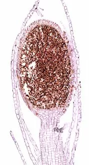
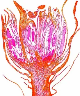
"Exploring the Intricate World of Plant Anatomy: A Fascinating Journey through Micrographs and Artwork" Delve into the hidden wonders with these captivating images. From dicotyledon plant stems to castor oil stems, each light micrograph unveils intricate structures that support life. Witness the mesmerizing beauty of Rhynchoglossum obliquum in artwork C016 / 5646, as its delicate details come to life. Dive beneath the surface with a glimpse at maize roots captured under a light microscope. Marvel at the complexity of English oak leaf pores revealed through scanning electron microscopy (SEM), showcasing nature's precision engineering. Take a closer look at the enchanting buttercup flower and tomato leaf, both magnified by light micrography. Explore pollination like never before, as SEM captures this pivotal moment in stunning detail. Uncover secrets hidden within French lavender leaf pores using SEM, revealing their unique structure and function. Witness gutation water droplets on dicot plants' leaf hairs through an unknown magnification SEM image - a phenomenon that showcases nature's ability to adapt and thrive. Embark on an exploration of Ladys mantle reproductive parts with SEM imagery, offering insights into reproduction strategies employed by these remarkable plants. Finally, get up close and personal with primrose pollen under SEM - tiny particles that play a crucial role in plant reproduction. Join us on this visual journey where science meets artistry; discover how every nook and cranny holds mysteries waiting to be unraveled in the fascinating world of plant anatomy.