
Plasma Cell Collection
"Unleashing the Power of Plasma Cells: A Visual Journey into Immune Response" In this captivating illustration

For sale as Licensed Images
Choose your image, Select your licence and Download the media
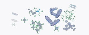
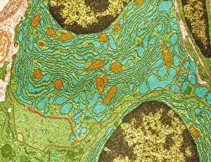
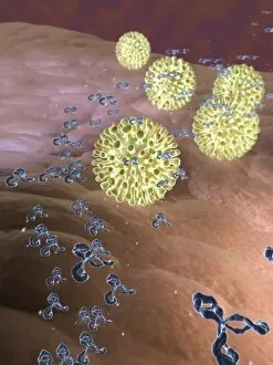
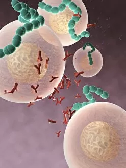
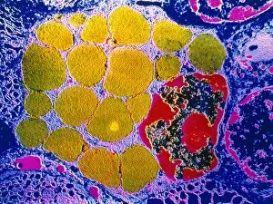
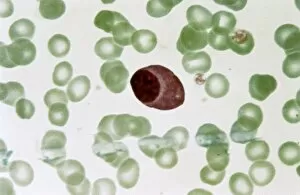
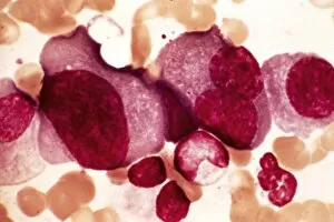
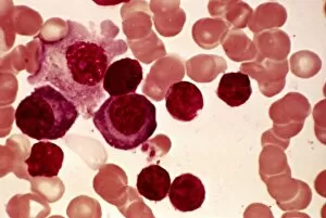
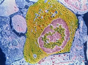
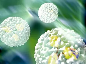
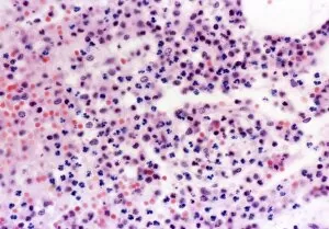
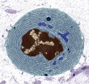
"Unleashing the Power of Plasma Cells: A Visual Journey into Immune Response" In this captivating illustration, we witness the remarkable response of plasma cells to an infection after vaccination. The scene unfolds with a microbe entering the body, triggering a cascade of events that will ultimately protect us from harm. As our immune system detects the presence of antigens on the invading microbe, it springs into action. Antibodies are produced by plasma cells, depicted here as vibrant warriors armed and ready to neutralize any threat. Their intricate structure is revealed through a high-resolution transmission electron microscope (TEM) image, showcasing their specialized role in combating infections. The artwork further explores how antibodies interact with different types of pathogens. In one frame, antibodies engage with viruses, while another showcases their battle against bacteria. These stunning visuals highlight the versatility and effectiveness of these tiny defenders in safeguarding our health. However, not all stories involving plasma cells have such positive outcomes. Multiple myeloma takes center stage in a light micrograph image capturing abnormal plasma cell proliferation within bone marrow. This glimpse into pathology reminds us that sometimes our own immune system can go awry. Returning to activated plasma cells under TEM magnification reveals their dynamic nature and highlights their critical role in mounting an effective defense against infections or other threats to our well-being. A second light micrograph offers insight into plasmocyte blood cells - close relatives to plasma cells - shedding light on their appearance within circulation and emphasizing their importance in maintaining immune homeostasis. Lastly, we delve into lymphoplasmacytic lymphoma through two distinct micrographs that illustrate its impact on lymphoid tissues. These images serve as poignant reminders that understanding diseases related to plasma cell dysfunction remains crucial for advancing medical knowledge and improving patient care. Through this visual journey exploring various aspects biology and pathology, we gain a deeper appreciation for these extraordinary cellular soldiers who tirelessly guard our bodies against harmful invaders while also reminding us of the complexities and challenges that can arise within our immune system.