
Polarised Light Micrograph Collection (#2)
Capturing the mesmerizing beauty of microscopic crystals through polarised light micrography is truly a sight to behold


For sale as Licensed Images
Choose your image, Select your licence and Download the media
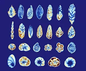
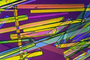
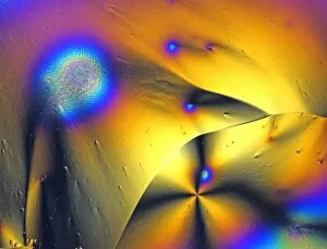
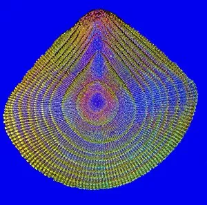
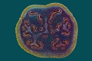
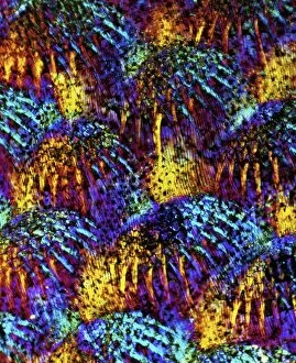
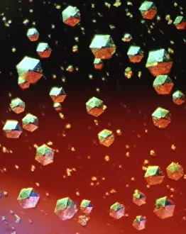
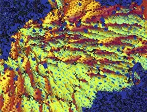
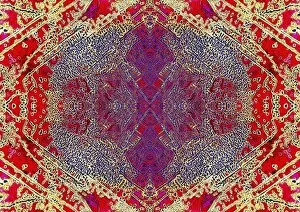
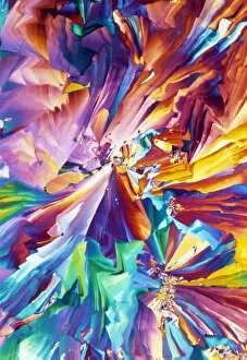
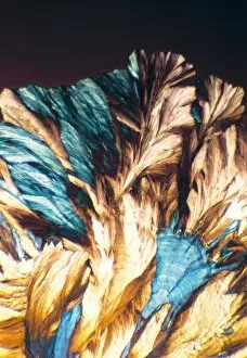
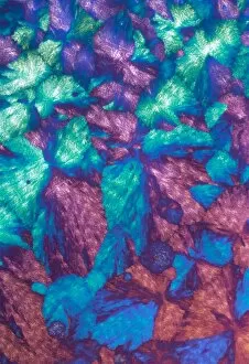
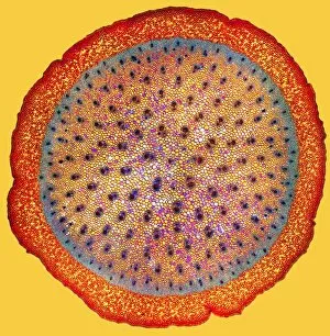
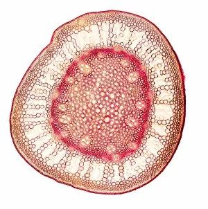
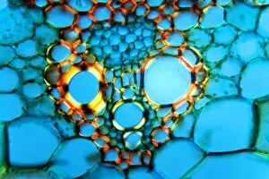
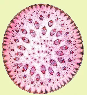
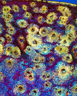
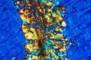
Capturing the mesmerizing beauty of microscopic crystals through polarised light micrography is truly a sight to behold. In this captivating world, caffeine crystals take center stage, showcasing their intricate patterns and delicate structure under the lens. The vibrant hues and symmetrical arrangements of these caffeine crystals create a visual spectacle that leaves us in awe. Moving on from caffeine, oxytocin hormone crystals make their appearance in PLM C016 / 7196, revealing an enchanting display of shimmering facets. These tiny wonders hold within them the power to evoke feelings of love and bonding. Similarly, oxytocin crystals captured in another light micrograph unveil their breathtaking elegance as they glisten with ethereal radiance. Cortisol crystals also join the lineup, offering a glimpse into their unique composition through yet another stunning light micrograph. Their sharp edges and intricate formations remind us of the complex nature of stress hormones present within our bodies. Returning to caffeine once again, its crystalline form takes on new dimensions when observed under polarised light microscopy. The interplay between colors and shapes creates an otherworldly landscape that transports us into a realm where science meets art. Testosterone makes its grand entrance through PLM crystal imagery, displaying its own distinct patterned arrangement that reflects strength and vitality. This snapshot allows us to appreciate the intricacies hidden within this essential hormone for both men and women alike. Insulin crystals come alive in yet another mesmerizing light micrograph (C017 / 8249), showcasing their delicate symmetry while reminding us of their vital role in regulating blood sugar levels within our bodies. Oxytocin hormone returns once more (LM C016 / 7195), captivating our imagination with its alluring crystal formation that hints at deep emotional connections we share as humans - bonds formed by trust, empathy, and compassion. Amidst these crystalline marvels lies a water fern rhizome captured under bright illumination.